Summary: Sulforaphane, a compound derived from broccoli sprouts, may be a useful new treatment for those suffering from schizophrenia. In a recent set of animal and human studies, researchers characterized novel chemical imbalances in the brain related to glutamate. Levels of glutamate, they discovered, can be altered by administering sulforaphane.
Source: Johns Hopkins Medicine
In a series of recently published studies using animals and people, Johns Hopkins Medicine researchers say they have further characterized a set of chemical imbalances in the brains of people with schizophrenia-related to the chemical glutamate. And they figured out how to tweak the level using a compound derived from broccoli sprouts.
They say the results advance the hope that supplementing with broccoli sprout extract, which contains high levels of the chemical sulforaphane, may someday provide a way to lower the doses of traditional antipsychotic medicines needed to manage schizophrenia symptoms, thus reducing unwanted side effects of the medicines.
“It’s possible that future studies could show sulforaphane to be a safe supplement to give people at risk of developing schizophrenia as a way to prevent, delay or blunt the onset of symptoms,” adds Akira Sawa, M.D., Ph.D., professor of psychiatry and behavioral sciences at the Johns Hopkins University School of Medicine and director of the Johns Hopkins Schizophrenia Center.
Schizophrenia is marked by hallucinations, delusions and disordered thinking, feeling, behavior, perception and speaking. Drugs used to treat schizophrenia don’t work completely for everyone, and they can cause a variety of undesirable side effects, including metabolic problems increasing cardiovascular risk, involuntary movements, restlessness, stiffness and “the shakes.”
In a study described in the Jan. 9 edition of the journal JAMA Psychiatry, the researchers looked for differences in brain metabolism between people with schizophrenia and healthy controls. They recruited 81 people from the Johns Hopkins Schizophrenia Center within 24 months of their first psychosis episode, which can be a characteristic symptom of schizophrenia, as well as 91 healthy controls from the community. The participants were an average of 22 years old, and 58% were men.
The researchers used a powerful magnet to measure and compare five regions in the brain between the people with and without psychosis. A computer analysis of 7-Tesla magnetic resonance spectroscopy (MRS) data identified individual chemical metabolites and their quantities.
The researchers found on average 4% significantly lower levels of the brain chemical glutamate in the anterior cingulate cortex region of the brain in people with psychosis compared to healthy people.
Glutamate is known for its role in sending messages between brain cells, and has been linked to depression and schizophrenia, so these findings added to evidence that glutamate levels have a role in schizophrenia.
Additionally, the researchers found a significant reduction of 3% of the chemical glutathione in the brain’s anterior cingulate cortex and 8% in the thalamus. Glutathione is made of three smaller molecules, and one of them is glutamate.
Next, the researchers asked how glutamate might be managed in the brain and whether that management is faulty in disease. They first looked at how it’s stored. Because glutamate is a building block of glutathione, the researchers wondered if the brain might use glutathione as a way to store extra glutamate. And if so, the researchers questioned if they could use known drugs to shift this balance to either release glutamate from storage when there isn’t enough, or send it into storage if there is too much.
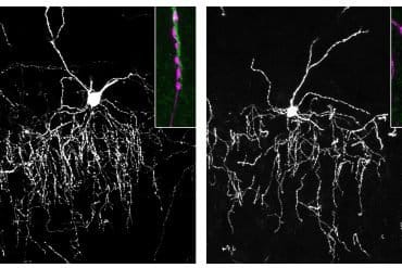
In another study, described in the Feb. 12 issue of the journal PNAS, the team used the drug L-Buthionine sulfoximine in rat brain cells to block an enzyme that turns glutamate into glutathione, allowing it to be used up. The researchers found that these nerves were more excited and fired faster, which means they were sending more messages to other brain cells. The researchers say shifting the balance this way is akin to shifting the brain cells to a pattern similar to one found in the brains of people with schizophrenia. Next, the researchers wanted to see if they could do the opposite and shift the balance to get more glutamate stored in the form of glutathione. They used the chemical sulforaphane found in broccoli sprouts, which is known to turn on a gene that makes more of the enzyme that sticks glutamate with another molecule to make glutathione. When they treated rat brain cells with glutathione, it slowed the speed at which the nerve cells fired, meaning they were sending fewer messages. The researchers say this pushed the brain cells to behave less like the pattern found in brains with schizophrenia.
“We are thinking of glutathione as glutamate stored in a gas tank,” says Thomas Sedlak, M.D., Ph.D., assistant professor of psychiatry and behavioral sciences. “If you have a bigger gas tank, you have more leeway on how far you can drive, but as soon as you take the gas out of the tank it’s burned up quickly. We can think of those with schizophrenia as having a smaller gas tank.”
Because sulforaphane changed the glutamate imbalance in the rat brains and affected how messages were transmitted between the rat brain cells, the researchers wanted to test whether sulforaphane could change glutathione levels in healthy people’s brains and see if this could eventually be a strategy for people with mental disorders. For their study, published in April 2018 in Molecular Neuropsychiatry, the researchers recruited nine healthy volunteers (four women, five men) to take two capsules with 100 micromoles daily of sulforaphane in the form of broccoli sprout extract for seven days.
The volunteers reported that a few of them were gassy and some had stomach upset when eating the capsules on an empty stomach, but overall the sulforaphane was relatively well tolerated.
The researchers used MRS again to monitor three brain regions for glutathione levels in the healthy volunteers before and after taking sulforaphane. They found that after seven days, there was about a 30% increase in average glutathione levels in the subjects’ brains. For example, in the hippocampus, glutathione levels rose an average of 0.27 millimolar from a baseline of 1.1 millimolar after seven days of taking sulforaphane.
The scientists say further research is needed to learn whether sulforaphane can safely reduce symptoms of psychosis or hallucinations in people with schizophrenia. They would need to determine an optimal dose and see how long people must take it to observe an effect. The researchers caution that their studies don’t justify or demonstrate the value of using commercially available sulforaphane supplements to treat or prevent schizophrenia, and patients should consult their physicians before trying any kind of over-the-counter supplement. Versions of sulforaphane supplements are sold in health food stores and at vitamin counters, and aren’t regulated by the U.S. Food and Drug Administration.

“For people predisposed to heart disease, we know that changes in diet and exercise can help stave off the disease, but there isn’t anything like that for severe mental disorders yet,” says Sedlak. “We are hoping that we will one day make some mental illness preventable to a certain extent.”
Sulforaphane is found in a variety of cruciferous vegetables and was first identified as a “chemoprotective” substance decades ago by Paul Talalay and Jed Fahey at Johns Hopkins.
According to the World Health Organization, schizophrenia affects about 21 million people worldwide.
Authors on the papers who were not yet mentioned include Anna Wang, Subechhya Pradhan, Jennifer Coughlin, Aditi Trivedi, Samantha DuBois, Jeffrey Crawford, Frederick Nucifora, Gerald Nestadt, Leslie Nucifora, David Schretlen, Peter Barker, Bindu Paul, Gregory Parker, Lynda Hester, Adele Snowman, Yu Taniguchi, Atsushi Kamiya, Solomon Snyder, Minori Koga, Lindsay Shaffer, Cecilia Higgs, Teppei Tanaka and Jed Fahey of Johns Hopkins.
Funding: These studies were supported by the National Institute of Mental Health (MH-094268, MH-092443, MH-084018, MH-105660, MH-107730, MH-096263 and U.S. Public Health Service MH18501), the National Institute of Biomedical Imaging and Bioengineering (EB015909), the National Institute of Neurological Disorders and Stroke (K08NS057824 and NS057824), the Stanley Foundation, the Stanley Research Foundation, the Johns Hopkins Brain Science Institute, the National Association for Research on Schizophrenia and Depression Young Investigator Grant and the Hilliard Fund. Participant recruitment was supported in part by Mitsubishi Tanabe Pharma.
Schretlen reported receiving royalties on sales of a test used in the JAMA Psychiatry paper under an agreement with Psychological Assessment Resources, which has been reviewed and approved by The Johns Hopkins University in accordance with its conflict of interest policies.
Source:
Johns Hopkins Medicine
Media Contacts:
Vanessa McMains – Johns Hopkins Medicine
Image Source:
The image is credited to Thomas Sedlak.
Original Research: Closed access
“Assessing Brain Metabolism With 7-T Proton Magnetic Resonance Spectroscopy in Patients With First-Episode Psychosis”. Anna M. Wang, PhD; Subechhya Pradhan, PhD; Jennifer M. Coughlin, MD; Aditi Trivedi, BS; Samantha L. DuBois, BA; Jeffrey L. Crawford, BA; Thomas W. Sedlak, MD, PhD; Fredrick C. Nucifora Jr, MD; Gerald Nestadt, MD; Leslie G. Nucifora, PhD; David J. Schretlen, PhD; Akira Sawa, MD, PhD; Peter B. Barker, DPhil.
JAMA Psychiatry. doi:10.1001/jamapsychiatry.2018.3637
Closed access
“The glutathione cycle shapes synaptic glutamate activity”. Thomas W. Sedlak, Bindu D. Paul, Gregory M. Parker, Lynda D. Hester, Adele M. Snowman, Yu Taniguchi, Atsushi Kamiya, Solomon H. Snyder, and Akira Sawa.
PNAS. doi:10.1073/pnas.1817885116
Open access
“Sulforaphane Augments Glutathione and Influences Brain Metabolites in Human Subjects: A Clinical Pilot Study”. Sedlak T.W., Nucifora L.G., Koga M., Shaffer L.S., Higgs C., Tanaka T., Wang A.M., Coughlin J.M., Barker P.B., Fahey J.W., and Sawa A.
Molecular Neuropsychiatry. doi:10.1159/000487639
Abstract
Assessing Brain Metabolism With 7-T Proton Magnetic Resonance Spectroscopy in Patients With First-Episode Psychosis
Importance The use of high-field magnetic resonance spectroscopy (MRS) in multiple brain regions of a large population of human participants facilitates in vivo study of localized or diffusely altered brain metabolites in patients with first-episode psychosis (FEP) compared to healthy participants.
Objective To compare metabolite levels in 5 brain regions between patients with FEP (evaluated within 2 years of onset) and healthy controls, and to explore possible associations between targeted metabolite levels and neuropsychological test performance.
Design, Setting, and Participants Cross-sectional design used 7-T MRS at a research MR imaging facility in participants recruited from clinics at the Johns Hopkins Schizophrenia Center and the local population. Eighty-one patients who had received a DSM-IV diagnosis of FEP within the last 2 years and 91 healthy age-matched (but not sex-matched) volunteers participated.
Main Outcomes and Measures Brain metabolite levels including glutamate, glutamine, γ-aminobutyric acid (GABA), N-acetylaspartate, N-acetylaspartyl glutamate, and glutathione, as well as performance on neuropsychological tests.
Results The mean (SD) age of 81 patients with FEP was 22.3 (4.4) years and 57 were male, while the mean (SD) age of 91 healthy participants was 23.3 (3.9) years and 42 were male. Compared with healthy participants, patients with FEP had lower levels of glutamate (F1,162 = 8.63, P = .02), N-acetylaspartate (F1,161 = 5.93, P = .03), GABA (F1,163 = 6.38, P = .03), and glutathione (F1,162 = 4.79, P = .04) in the anterior cingulate (all P values are corrected for multiple comparisons); lower levels of N-acetylaspartate in the orbitofrontal region (F1,136 = 7.23, P = .05) and thalamus (F1,133 = 6.78, P = .03); and lower levels of glutathione in the thalamus (F1,135 = 7.57, P = .03). Among patients with FEP, N-acetylaspartate levels in the centrum semiovale white matter were significantly correlated with performance on neuropsychological tests, including processing speed (r = 0.48; P < .001), visual (r = 0.33; P = .04) and working (r = 0.38; P = .01) memory, and overall cognitive performance (r = 0.38; P = .01).
Conclusions and Relevance Seven-tesla MRS offers insights into biochemical changes associated with FEP and may be a useful tool for probing brain metabolism that ranges from neurotransmission to stress-associated pathways in participants with psychosis.
Abstract
The glutathione cycle shapes synaptic glutamate activity
Glutamate is the most abundant excitatory neurotransmitter, present at the bulk of cortical synapses, and participating in many physiologic and pathologic processes ranging from learning and memory to stroke. The tripeptide, glutathione, is one-third glutamate and present at up to low millimolar intracellular concentrations in brain, mediating antioxidant defenses and drug detoxification. Because of the substantial amounts of brain glutathione and its rapid turnover under homeostatic control, we hypothesized that glutathione is a relevant reservoir of glutamate and could influence synaptic excitability. We find that drugs that inhibit generation of glutamate by the glutathione cycle elicit decreases in cytosolic glutamate and decreased miniature excitatory postsynaptic potential (mEPSC) frequency. In contrast, pharmacologically decreasing the biosynthesis of glutathione leads to increases in cytosolic glutamate and enhanced mEPSC frequency. The glutathione cycle can compensate for decreased excitatory neurotransmission when the glutamate-glutamine shuttle is inhibited. Glutathione may be a physiologic reservoir of glutamate neurotransmitter.
Abstract
Sulforaphane Augments Glutathione and Influences Brain Metabolites in Human Subjects: A Clinical Pilot Study
Schizophrenia and other neuropsychiatric disorders await mechanism-associated interventions. Excess oxidative stress is increasingly appreciated to participate in the pathophysiology of brain disorders, and decreases in the major antioxidant, glutathione (GSH), have been reported in multiple studies. Technical cautions regarding the estimation of oxidative stress-related changes in the brain via imaging techniques have led investigators to explore peripheral GSH as a possible pathological signature of oxidative stress-associated brain changes. In a preclinical model of GSH deficiency, we found a correlation between whole brain and peripheral GSH levels. We found that the naturally occurring isothiocyanate sulforaphane increased blood GSH levels in healthy human subjects following 7 days of daily oral administration. In parallel, we explored the potential influence of sulforaphane on brain GSH levels in the anterior cingulate cortex, hippocampus, and thalamus via 7-T magnetic resonance spectroscopy. A significant positive correlation between blood and thalamic GSH post- and pre-sulforaphane treatment ratios was observed, in addition to a consistent increase in brain GSH levels in response to treatment. This clinical pilot study suggests the value of exploring relationships between peripheral GSH and clinical/neuropsychological measures, as well as the influences sulforaphane has on functional measures that are altered in neuropsychiatric disorders.


