

Summary: In adolescents, brain areas associated with emotional, social, and cognitive functions appear to remain more plastic, or malleable, than other brain areas, thus rendering young people more sensitive to socioeconomic environments throughout adolescence.
Source: University of Pennsylvania
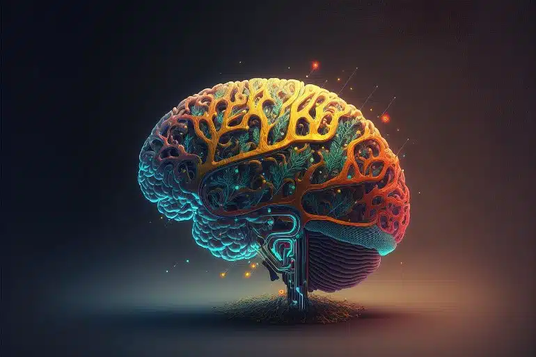
Brain development does not occur uniformly across the brain, but follows a newly identified developmental sequence, according to a new Penn Medicine study.
Brain regions that support cognitive, social, and emotional functions appear to remain malleable—or capable of changing, adapting, and remodeling—longer than other brain regions, rendering youth sensitive to socioeconomic environments through adolescence.
The findings were published recently in Nature Neuroscience.
Researchers charted how developmental processes unfold across the human brain from the ages of 8 to 23 years old through magnetic resonance imaging (MRI). The findings indicate a new approach to understanding the order in which individual brain regions show reductions in plasticity during development.
Brain plasticity refers to the capacity for neural circuits—connections and pathways in the brain for thought, emotion, and movement—to change or reorganize in response to internal biological signals or the external environment. While it is generally understood that children have higher brain plasticity than adults, this study provides new insights into where and when reductions in plasticity occur in the brain throughout childhood and adolescence.
The findings reveal that reductions in brain plasticity occur earliest in “sensory-motor” regions, such as visual and auditory regions, and occur later in “associative” regions, such as those involved in higher-order thinking (problem solving and social learning). As a result, brain regions that support executive, social, and emotional functions appear to be particularly malleable and responsive to the environment during early adolescence, as plasticity occurs later in development.
“Studying brain development in the living human brain is challenging. A lot of neuroscientists’ understanding about brain plasticity during development actually comes from studies conducted with rodents. But rodent brains do not have many of what we refer to as the association regions of the human brain, so we know less about how these important areas develop,” said corresponding author Theodore D. Satterthwaite, MD, the McLure Associate Professor of Psychiatry in the Perelman School of Medicine at the University of Pennsylvania, and director of the Penn Lifespan Informatics and Neuroimaging Center (PennLINC).
To address this challenge, the researchers focused on comparing insights from previous rodent studies to youth MRI imaging insights. Prior research examining how neural circuits behave when they are plastic uncovered that brain plasticity is linked to a unique pattern of “intrinsic” brain activity. Intrinsic activity is the neural activity occurring in a part of the brain when it is at rest, or not being engaged by external stimuli or a mental task.
When a brain region is less developed and more plastic, there tends to be more intrinsic activity within the region, and that activity also tends to be more synchronized. This is because more neurons in the region are active, and they tend to be active at the same time. As a result, measurements of brain activity waves show an increase in amplitude (or height).
“Imagine that individual neurons within a region of the brain are like instruments in an orchestra. As more instruments begin to play together in synchrony, the sound level of the orchestra increases, and the amplitude of the sound wave gets higher,” said first author Valerie Sydnor, a Neuroscience PhD student.
“Just like decibel meters can measure the amplitude of a sound wave, the amplitude of intrinsic brain activity can be measured with functional MRI while kids are simply resting in the scanner. This allowed our team to study a functional marker of brain plasticity safely and non-invasively in youth.”
Analyzing MRI scans from more than 1,000 individuals, the authors found that the functional marker of brain plasticity declined in earlier childhood in sensory-motor regions but did not decline until mid-adolescence in associative regions.
“These slow-developing associative regions are also those that are vital for children’s cognitive attainment, social interactions, and emotional well-being,” Satterthwaite added. “We are really starting to understand the uniqueness of human’s prolonged developmental program.”
“If a brain region remains malleable for longer, it may also remain sensitive to environmental influences for a longer window of development,” Sydnor said. “This study found evidence for just that.”
The authors studied relationships between youths’ socioeconomic environments and the same functional marker of plasticity. They found that the effects of the environment on the brain were not uniform across regions nor static across development. Rather, the effects of the environment on the brain changed as the identified developmental sequence progressed.
Critically, youths’ socioeconomic environments generally had a larger impact on brain development in the late-maturing associative brain regions, and the impact was found to be largest in adolescence.
“This work lays the foundation for understanding how the environment shapes neurodevelopmental trajectories even through the teenage years,” said Bart Larsen, PhD, a PennLINC postdoctoral researcher and co-author.
Sydnor elaborated, “The hope is that studying developmental plasticity will help us to understand when environmental enrichment programs will have a beneficial impact on each child’s neurodevelopmental trajectory. Our findings support that programs designed to alleviate disparities in youths’ socioeconomic environments remain important for brain development throughout the adolescent period.”
Funding: This study was supported by the National Institute of Health (R01MH113550, R01MH120482, R01MH112847, R01MH119219, R01MH123563, R01MH119185, R01MH120174, R01NS060910, R01EB022573, RF1MH116920., RF1MH121867, R37MH125829, R34DA050297, K08MH120564, K99MH127293, T32MH014654). The study was also supported by the National Science Foundation Graduate Research Fellowship (DGE-1845298).
Additional support was provided by the Penn-CHOP Lifespan Brain Institute and the Penn Center for Biomedical Image Computing and Analytics.
About this brain plasticity research news
Author: Eric Horvath
Source: University of Pennsylvania
Contact: Eric Horvath – University of Pennsylvania
Image: The image is in the public domain
Original Research: Closed access.
“Intrinsic activity development unfolds along a sensorimotor–association cortical axis in youth” by Valerie Sydnor et al. Nature Neuroscience
Abstract
Intrinsic activity development unfolds along a sensorimotor–association cortical axis in youth
Animal studies of neurodevelopment have shown that recordings of intrinsic cortical activity evolve from synchronized and high amplitude to sparse and low amplitude as plasticity declines and the cortex matures.
Leveraging resting-state functional MRI (fMRI) data from 1,033 youths (ages 8–23 years), we find that this stereotyped refinement of intrinsic activity occurs during human development and provides evidence for a cortical gradient of neurodevelopmental change.
Declines in the amplitude of intrinsic fMRI activity were initiated heterochronously across regions and were coupled to the maturation of intracortical myelin, a developmental plasticity regulator. Spatiotemporal variability in regional developmental trajectories was organized along a hierarchical, sensorimotor–association cortical axis from ages 8 to 18.
The sensorimotor–association axis furthermore captured variation in associations between youths’ neighborhood environments and intrinsic fMRI activity; associations suggest that the effects of environmental disadvantage on the maturing brain diverge most across this axis during midadolescence.
These results uncover a hierarchical neurodevelopmental axis and offer insight into the progression of cortical plasticity in humans.


