Summary: A new technology that allows researchers to examine circulation in the brain could help to identify early signs of neurological problems.
Source: University of Surrey.
A study published today in the Journal of Anatomy has made an important breakthrough in the examination of blood vessels in the brain giving scientists a clearer understanding of how dementia, brain cancer and stroke can affect veins and capillaries in this organ.
Working collaboratively researchers from the the School of Veterinary Medicine at the University of Surrey and the Federal University of Sao Paulo developed an innovative technique to examine and quantify blood vessels in the brain using 3D Image Analysis (Stereology) procedures.
Using experimental animal models, this technique will allow scientists to study how such diseases develop in the brain and help them identify, through examination of blood vessels, potential warning signs of illnesses before symptoms appear. These learnings can potentially be translated into humans and help reduce the number of deaths from these illnesses. The procedure can also be used in post mortems and biopsies examinations of animal and human tissue making it easier for pathologists to determine causes of death and quickly identify alterations in the brain circulation (such as clots) or tumors.
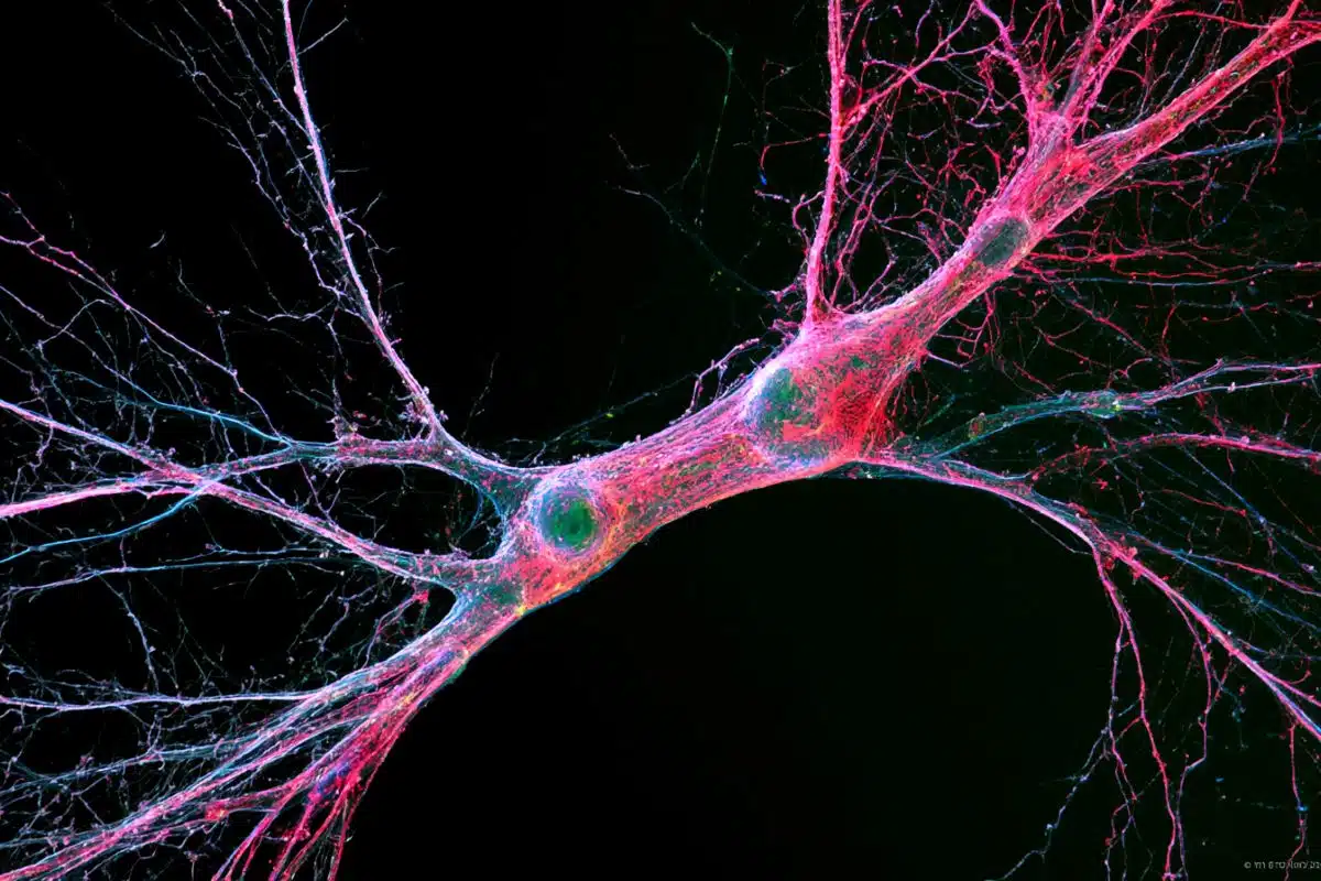
The inexpensive technique of dissolving China Ink with gelatin creates a solution making blood vessels more visible with the use of a confocal microscope. This enables scientists and pathologists to make an accurate reading of their number, length, surface area and create 3D images which can help identify changes in their shape and size, key indicators of a number of circulation-related diseases of the brain.
This innovative method will also facilitate a greater understanding of how exercise affects the brain. Scientists will now be able to examine circulatory effects of increased or decreased heart rate, arterial pressure on the brain and the creation of new vessels (angiogenesis).

Co-author of the study Dr Augusto Coppi from the University of Surrey said: “The brain is a fascinating organ but our full understanding of its circulation is lacking. Previously we have been unable to fully sample and perform a quantification of the circulation of the brain in 3D as we simply could not see all vessels due to their minute size and sometimes due to their irregular spatial distribution.
“This new technique will allow us to sample, image and count blood vessels in 3D giving us a greater mechanistic comprehension of how the circulation of the brain works and how brain diseases such as dementia and stroke affect this organ. With an estimated 850,000 people diagnosed with dementia in England, this technique marks a significant breakthrough in the fight against this disease.”
Source: Peter La – University of Surrey
Image Source: NeuroscienceNews.com image is adapted from the University of Surrey press release.
Original Research: Abstract for “Reflection imaging of China ink-perfused brain vasculature using confocal laser-scanning microscopy after clarification of brain tissue by the Spalteholz method” by R. C. Gutierre, D. Vannucci Campos, R. A. Mortara, A. A. Coppi, and R. M. Arida in Journal of Anatomy. Published online December 2016 doi:10.1111/joa.12578
[cbtabs][cbtab title=”MLA”]University of Surrey “Examining Blood Vessels in 3D to Unlock Secrets of the Brain.” NeuroscienceNews. NeuroscienceNews, 7 January 2017.
<https://neurosciencenews.com/3d-blood-vessel-brain-5887/>.[/cbtab][cbtab title=”APA”]University of Surrey (2017, January 7). Examining Blood Vessels in 3D to Unlock Secrets of the Brain. NeuroscienceNew. Retrieved January 7, 2017 from https://neurosciencenews.com/3d-blood-vessel-brain-5887/[/cbtab][cbtab title=”Chicago”]University of Surrey “Examining Blood Vessels in 3D to Unlock Secrets of the Brain.” https://neurosciencenews.com/3d-blood-vessel-brain-5887/ (accessed January 7, 2017).[/cbtab][/cbtabs]
Abstract
Reflection imaging of China ink-perfused brain vasculature using confocal laser-scanning microscopy after clarification of brain tissue by the Spalteholz method
Confocal laser-scanning microscopy is a useful tool for visualizing neurons and glia in transparent preparations of brain tissue from laboratory animals. Currently, imaging capillaries and venules in transparent brain tissues requires the use of fluorescent proteins. Here, we show that vessels can be imaged by confocal laser-scanning microscopy in transparent cortical, hippocampal and cerebellar preparations after clarification of China ink-injected specimens by the Spalteholz method. This method may be suitable for global, three-dimensional, quantitative analyses of vessels, including stereological estimations of total volume and length and of surface area of vessels, which constitute indirect approaches to investigate angiogenesis.
“Reflection imaging of China ink-perfused brain vasculature using confocal laser-scanning microscopy after clarification of brain tissue by the Spalteholz method” by R. C. Gutierre, D. Vannucci Campos, R. A. Mortara, A. A. Coppi, and R. M. Arida in Journal of Anatomy. Published online December 2016 doi:10.1111/joa.12578