Summary: Researchers have released data from the world’s largest brain and body scanning study.
Source: Imperial College London.
Data from the world’s largest brain and body scanning study has been released.
Exciting early results from analysing the brain imaging data, alongside thousands of measures of lifestyle, physical fitness, cognitive health and physical measures such as body-mass-index (BMI) and bone density have been published in Nature Neuroscience.
The high quality of the imaging data and very large number of subjects allowed researchers to identify more than 30,000 significant associations between the many different brain imaging measures and the non-imaging measures. The findings have now been made available for use by researchers worldwide.
Results included:
- Strong associations between people’s cognitive processing speed and markers of the integrity of the brain’s “wiring” and the size of brain structures. These effects increased in strength as people aged.
- A negative correlation between brain activity during a simple shape-matching task and intelligence, an effect that didn’t relate to participants’ age. This might be because the people who scored more highly on the cognitive tests needed to use less of their brain to carry out the task.
- A pattern of strong associations between higher blood pressure, greater alcohol consumption, and several measures that could reflect injury to connections in the brain.
- A separate pattern of correlations, linking intake of alcohol and tobacco and changes in red blood cells and cardiac fitness, to brain imaging signals associated with increased iron deposits in the brain.
- Researchers also unearthed some more complicated patterns of correlation. For example, one pattern links brain imaging to intelligence, level of education, and a set of lifestyle factors that at first appear unrelated – including amount of time spent outdoors.
- It is plausible that, taken together, these factors create a profile of socio-economic-status and its relation to the brain.
- However, because UK Biobank is an “observational” study that characterizes a cross-section of individuals, it’s not always straightforward to establish which factors cause which, but such results should help scientists to define much more precise questions to address in the future search for ways of preventing or treating brain disease.
UK Biobank will be the world’s largest health imaging study. The imaging is funded by the Medical Research Council, Wellcome Trust, and the British Heart Foundation. It was launched in April 2016 after a number of years of planning and consultation with a large number of health and scanning experts. With the ambitious goal of imaging 100,000 existing UK Biobank participants, it is creating the biggest collection of scans of internal organs, to transform the way scientists study a wide range of diseases, including dementia, arthritis, cancer, heart attacks and stroke.
Today’s paper describes the brain imaging part of UK Biobank, led by Professsors Steve Smith and Karla Miller from the University of Oxford, and Professor Paul Matthews from Imperial College London.
Professor Miller said: “We are using cutting-edge MRI scans and Big Data analysis methods to get the most comprehensive window into the brain that current imaging technology allows.”
“These results are just a first glimpse into this massive, rich dataset will that emerge in the coming years. It is an unparalleled resource that will transform our understanding of many common diseases.”
Professor Matthews, Edmond and Lily Safra Chair and Head of Brain Sciences at Imperial, added: “These results are exciting, but merely provide a first hint of what can be discovered with the UK Biobank. This project also is a landmark because of the way it has been done: 500,000 volunteers across the U.K. are donating their time to be part of it and more than 125 scientists from across the world contributed to the design of the imaging enhancement alone. Imperial College scientists played a major role in its inception and leadership as part of a team recruited by the U.K. biobank from a number of UK universities. This is a wonderful example of “open science”.
The paper reports first results from this remarkable data resource, which includes six different kinds of brain imaging done in the 30 minutes that each volunteer is in the brain scanner.
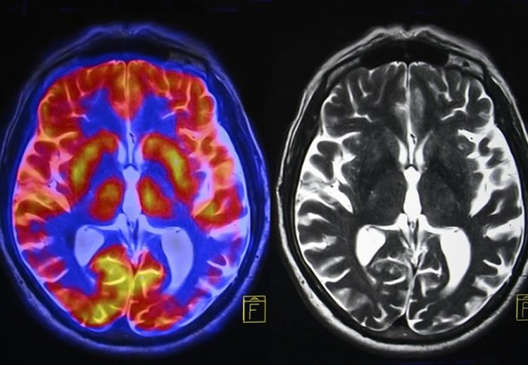
Professor Smith explained: “We have ‘structural imaging’ – that tells us about brain anatomy – the shapes and sizes of the different parts of the brain. Another kind – ‘functional MRI’ – tells us about complex patterns of brain activity. Yet another kind – ‘diffusion MRI’ – tells us about the brain’s wiring diagram. The rich and diverse information contained in these scans will reveal how the working of the brain can change with aging and disease; different diseases will best be understood through different combinations of information across these different images.”
UK Biobank has already scanned 10,000 participants, including images of the heart, body, bone and blood vessels in addition to brain scans. This will be by far the largest brain imaging study ever conducted; within another 5 years UK Biobank will have completed the scanning of 100,000 participants.
One reason for needing such large numbers of participants is to have enough subjects to allow discovery of early, possibly subtle, markers of future disease risk, both for a range of common diseases and for rare neurological disorders like motor neuron disease.
An important objective of the UK Biobank is to provide a resource for discovery of new insights into diseases like Alzheimer’s, which demands scanning healthy subjects years or decades before they develop symptoms. From the UK Biobank data, scientists anywhere can aim to learn much more about brain diseases – and their relationship to a broad range of other diseases or disease risks – to guide the development of earlier targeted treatment (or changes in lifestyle) that could in the future prevent major diseases from ever happening.
Source: Kate Wighton – Imperial College London
Image Source: This NeuroscienceNews.com image is adapted from the Imperial College London press release.
Original Research: Abstract for “Multimodal population brain imaging in the UK Biobank prospective epidemiological study” by Karla L Miller, Fidel Alfaro-Almagro, Neal K Bangerter, David L Thomas, Essa Yacoub, Junqian Xu, Andreas J Bartsch, Saad Jbabdi, Stamatios N Sotiropoulos, Jesper L R Andersson, Ludovica Griffanti, Gwenaëlle Douaud, Thomas W Okell, Peter Weale, Iulius Dragonu, Steve Garratt, Sarah Hudson, Rory Collins, Mark Jenkinson, Paul M Matthews and Stephen M Smith in Nature Neuroscience. Published online September 19 2016 doi:10.1038/nn.4393
[cbtabs][cbtab title=”MLA”]Imperial College London. “Alcohol, Tobacco and Time Spent Outdoors Linked to Brain Connections.” NeuroscienceNews. NeuroscienceNews, 20 September 2016.
<https://neurosciencenews.com/tobacco-alcohol-lifestyle-neural-network-5082/>.[/cbtab][cbtab title=”APA”]Imperial College London. (2016, September 20). Alcohol, Tobacco and Time Spent Outdoors Linked to Brain Connections. NeuroscienceNews. Retrieved September 20, 2016 from https://neurosciencenews.com/tobacco-alcohol-lifestyle-neural-network-5082/[/cbtab][cbtab title=”Chicago”]Imperial College London. “Alcohol, Tobacco and Time Spent Outdoors Linked to Brain Connections.” https://neurosciencenews.com/tobacco-alcohol-lifestyle-neural-network-5082/ (accessed September 20, 2016).[/cbtab][/cbtabs]
Abstract
Multimodal population brain imaging in the UK Biobank prospective epidemiological study
Medical imaging has enormous potential for early disease prediction, but is impeded by the difficulty and expense of acquiring data sets before symptom onset. UK Biobank aims to address this problem directly by acquiring high-quality, consistently acquired imaging data from 100,000 predominantly healthy participants, with health outcomes being tracked over the coming decades. The brain imaging includes structural, diffusion and functional modalities. Along with body and cardiac imaging, genetics, lifestyle measures, biological phenotyping and health records, this imaging is expected to enable discovery of imaging markers of a broad range of diseases at their earliest stages, as well as provide unique insight into disease mechanisms. We describe UK Biobank brain imaging and present results derived from the first 5,000 participants’ data release. Although this covers just 5% of the ultimate cohort, it has already yielded a rich range of associations between brain imaging and other measures collected by UK Biobank.
“Multimodal population brain imaging in the UK Biobank prospective epidemiological study” by Karla L Miller, Fidel Alfaro-Almagro, Neal K Bangerter, David L Thomas, Essa Yacoub, Junqian Xu, Andreas J Bartsch, Saad Jbabdi, Stamatios N Sotiropoulos, Jesper L R Andersson, Ludovica Griffanti, Gwenaëlle Douaud, Thomas W Okell, Peter Weale, Iulius Dragonu, Steve Garratt, Sarah Hudson, Rory Collins, Mark Jenkinson, Paul M Matthews and Stephen M Smith in Nature Neuroscience. Published online September 19 2016 doi:10.1038/nn.4393