Summary: Scripps researchers have uncovered the process that helps control neuron growth.
Source: Scripps Research Institute.
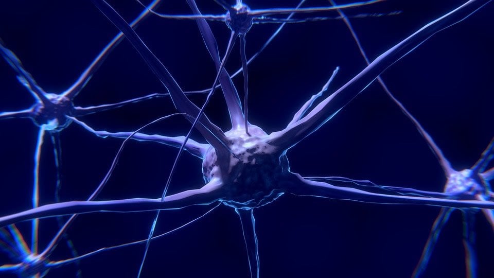
All things must come to an end. This is particularly true for neurons, especially the extensions called axons that transmit electrochemical signals to other nerve cells. Without controlled termination of individual neuron growth, the efficient and accurate construction of a nervous system is in serious jeopardy.
Scientists from the Florida campus of The Scripps Research Institute (TSRI) have now uncovered new insights into the regulatory network behind that termination. The study, led by TSRI Associate Professor Brock Grill, PhD, was recently published online ahead of print in the journal Development.
The scientists focused on axons, long cellular structures that project outward from the neuron body. When nerve cells fire, it’s the axon that transmits the electrochemical signal to other neurons. Over the course of their development, axons extend, change their growth in response to cellular guidance cues and form synapses.
At the heart of this process is a specialized structure on the end of each axon called a growth cone. Successful development depends on the growth cone stopping at the correct destination and when the axon is the correct length, a process known as axon termination.
Using the nematode worm C. elegans as a model, Grill and his colleagues found for the first time that growth cone collapse prior to axon termination is protracted as the growth cone transitions from a dynamic to a static state.
“We know very little about the process of how axons actually stop growing in a living animal,” Grill says. “What we found in our simple, but powerful model is that a signaling hub protein called RPM-1 is required to regulate the collapse of growth cones during axon termination.”
It’s the protracted nature of the process, Grill says, that is likely to make the transition-and the termination-permanent.
These findings provide new details on how growth cone collapse is regulated during axon termination in vivo. The study also shows that RPM-1 signaling destabilizes nerve cell microtubules-large molecules that provide critical cell structure-to facilitate growth cone collapse and axon termination.
When the scientists looked at the relationship between RPM-1 and other regulators of microtubule stability, they were surprised by the results.
They found that that while RPM-1 signaling destabilizes axon microtubules, the microtubule stabilizer Tau potentially inhibits RPM -1, something that was previously unknown. “People have very little knowledge about how TAU works under normal physiological conditions,” says TSRI Research Associate Melissa Borgen, PhD, first author of the study.
“Our results suggest that Tau inhibition of RPM-1 is necessary for proper axon development, and offers the first evidence that RPM-1 can be regulated in vivo in neurons.”
The research has implications for the development of neurological disorders as well. In mouse models, RPM-1 is an active force in axon degeneration and TAU has been linked to neurological disorders, including Alzheimer’s disease and frontal temporal dementia.
“You wouldn’t necessarily have thought Tau and RPM-1 would function this way,” Grill says. “That’s the power of genetics. Although we assessed the genetic relationship between Tau and RPM-1 in axon development, our results could have important implications for neurodegeneration.”
In addition to Grill and Borgen, the other author of the study, “RPM-1 Regulates Axon Termination by Affecting Growth Cone Collapse and Microtubule Stability,” is Dandan Wang, PhD, of TSRI.
Funding: The study was supported by the National Institutes of Health (grant R01 NS072129) and the National Science Foundation (grant IOS-1121095).
Source: David Barnstone – Scripps Research Institute
Publisher: Organized by NeuroscienceNews.com.
Image Source: NeuroscienceNews.com image is in the public domain.
Original Research: Abstract for “RPM-1 regulates axon termination by affecting growth cone collapse and microtubule stability” by Melissa A. Borgen, Dandan Wang, and Brock Grill in Development. Published online December 2017 doi:10.1242/dev.154187
[cbtabs][cbtab title=”MLA”]Scripps Research Institute “‘Simple, But Powerful’ Model Reveals Mechanism Behind Neuron Development.” NeuroscienceNews. NeuroscienceNews, 18 December 2017.
<https://neurosciencenews.com/neuron-development-model-8204/>.[/cbtab][cbtab title=”APA”]Scripps Research Institute (2017, December 18). ‘Simple, But Powerful’ Model Reveals Mechanism Behind Neuron Development. NeuroscienceNews. Retrieved December 18, 2017 from https://neurosciencenews.com/neuron-development-model-8204/[/cbtab][cbtab title=”Chicago”]Scripps Research Institute “‘Simple, But Powerful’ Model Reveals Mechanism Behind Neuron Development.” https://neurosciencenews.com/neuron-development-model-8204/ (accessed December 18, 2017).[/cbtab][/cbtabs]
Abstract
RPM-1 regulates axon termination by affecting growth cone collapse and microtubule stability
Axon termination is essential for efficient and accurate nervous system construction. At present, relatively little is known about how growth cone collapse occurs prior to axon termination in vivo. Using the mechanosensory neurons of C. elegans, we found collapse prior to axon termination is protracted, with the growth cone transitioning from a dynamic to a static state. Growth cone collapse prior to termination is facilitated by the signaling hub RPM-1. Given the prominence of the cytoskeleton in growth cone collapse, we assessed the relationship between RPM-1 and regulators of actin dynamics and microtubule stability. Our results reveal several important findings about how axon termination is orchestrated: (1) RPM-1 functions in parallel to RHO-1 and CRMP/UNC-33, but is suppressed by the Rac isoform MIG-2; (2) RPM-1 opposes the function of microtubule stabilizers, including tubulin acetyltransferases; and (3) genetic epistasis suggests the microtubule-stabilizing protein Tau/PTL-1 potentially inhibits RPM-1. These findings provide insight into how growth cone collapse is regulated during axon termination in vivo, and suggest that RPM-1 signaling destabilizes microtubules to facilitate growth cone collapse and axon termination.
“RPM-1 regulates axon termination by affecting growth cone collapse and microtubule stability” by Melissa A. Borgen, Dandan Wang, and Brock Grill in Development. Published online December 2017 doi:10.1242/dev.154187