Summary: A new study explores the role microtubles play in neurodevelopment.
Source: Drexel University.
With a boost from motor proteins, sliding microtubules give nerve cells a smoother ride.
As the human brain develops, neurons leave their birthplace and take a trip to distant locations. Once they reach their final destination, the neurons then send out axons and dendrites — the branches that receive and send messages from other cells.
Humans’ most basic functions depend on this journey of neurons getting to where they need to go, and making correct connections once they arrive. This ensures that our eyes can see, our ears can hear, our fingers can touch and so on.
A new study from Drexel University researchers shows that the sliding movements of a small group of intracellular structures — called microtubules — play a key role in keeping neurons on a smooth, proper trajectory.
This discovery could ultimately help researchers better understand how neurons gone astray contribute to neurodevelopmental disorders, said Peter Baas, PhD, a professor in the College of Medicine and the study’s principal investigator.
“This study is important for understanding how a healthy brain is organized,” Baas said. “If neurons do not know when to start migrating, or where to go, or if the axons don’t grow long enough, that sort of thing can give way to disorders such as autism.”
The study, published this month in the Journal of Cell Biology, focuses on microtubules and the molecular motor proteins that generate forces on these intracellular structures.
Until recently, the primary school of thought accepted that microtubules’ main functions were to grow longer and shorter — as merely “passive players” in the wiring of the nervous system. However, Baas has spent his career studying the ways in which motor proteins push and pull on microtubules, causing them turn, and thus enabling the axon to move in response to cues inside the embryo.
Researchers also assumed that all “functionally relevant” microtubules were attached to the centrosome, the main organization center of a cell, according to Baas.
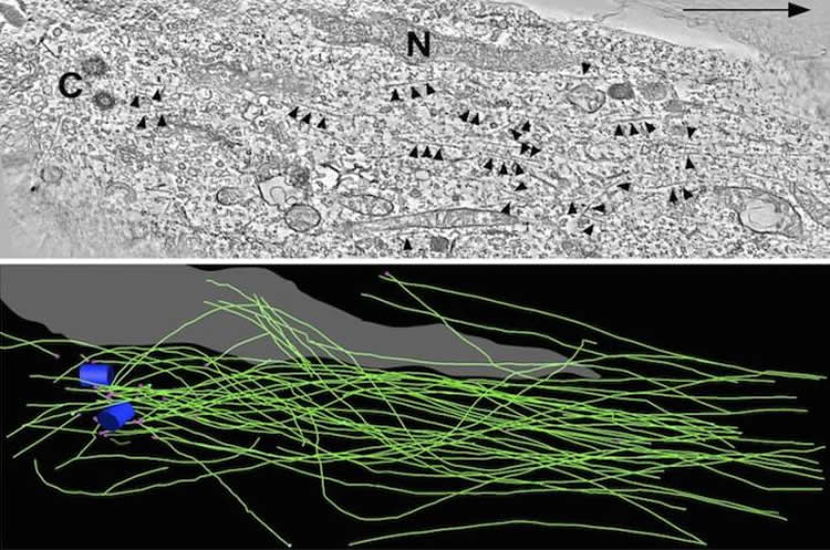
Baas and his research team decided to use electron tomography — the most rigorous imaging method available — to see, first, whether some microtubules might actually be detached from the centrosome, and if so, how that detachment might contribute to neuron migration.
The researchers found that a small group of microtubules were not attached to the centrosome, and that motor proteins can actually slide these unattached microtubules within the neuron as it migrates.
Next, they wanted to know, do those sliding, unattached microtubules matter?
To find out, the researchers added a drug to immobilize them. They saw that, although the neurons still moved, they frequently changed direction, instead of migrating in a simple, straight line.
“When we used the drug that inhibits sliding, we saw that the neuron can’t migrate in a nice straight, smooth trajectory,” Baas said. “That’s how we found out that little bit of sliding that normally occurs is really important for maneuverability.”
Going a step further, the researchers detached more microtubules from the centrosome by knocking out their anchoring protein. This caused many of the neurons to slow down or even come to a complete halt, and the neurons’ axons continued to grow at long lengths.
By manipulating levels of protein, the researchers now know that even the smallest alterations can greatly change the morphology and migratory behavior of a neuron, which can translate to developmental problems.
“If any of these mechanisms – with ninein or any of these motor proteins — are disrupted, there can be problems anywhere along the way,” Baas said.
Funding: The work was supported by the Craig H. Neilsen Foundation, NIH/National Institute of Neurological Disorders and Stroke), US Department of Defense.
Source: Lauren Ingeno – Drexel University
Image Source: This NeuroscienceNews.com image is credited to Peter Baas.
Original Research: Abstract for “Sliding of centrosome-unattached microtubules defines key features of neuronal phenotype” by Anand N. Rao, Aditi Falnikar, Eileen T. O’Toole, Mary K. Morphew, Andreas Hoenger, Michael W. Davidson, Xiaobing Yuan, and Peter W. Baas in Journal of Cell Biology. Published online May 2 2016 doi:10.1083/jcb.201506140
[cbtabs][cbtab title=”MLA”]Drexel University. “How Neurons Reach Their Final Destination.” NeuroscienceNews. NeuroscienceNews, 17 May 2016.
<https://neurosciencenews.com/microtubules-axons-neurons-4242/>.[/cbtab][cbtab title=”APA”]Drexel University. (2016, May 17). How Neurons Reach Their Final Destination. NeuroscienceNews. Retrieved May 17, 2016 from https://neurosciencenews.com/microtubules-axons-neurons-4242/[/cbtab][cbtab title=”Chicago”]Drexel University. “How Neurons Reach Their Final Destination.” NeuroscienceNews.
https://neurosciencenews.com/microtubules-axons-neurons-4242/ (accessed May 17, 2016).[/cbtab][/cbtabs]
Abstract
Sliding of centrosome-unattached microtubules defines key features of neuronal phenotype
Contemporary models for neuronal migration are grounded in the view that virtually all functionally relevant microtubules (MTs) in migrating neurons are attached to the centrosome, which occupies a position between the nucleus and a short leading process. It is assumed that MTs do not undergo independent movements but rather transduce forces that enable movements of the centrosome and nucleus. The present results demonstrate that although this is mostly true, a small fraction of the MTs are centrosome-unattached, and this permits limited sliding of MTs. When this sliding is pharmacologically inhibited, the leading process becomes shorter, migration of the neuron deviates from its normal path, and the MTs within the leading process become buckled. Partial depletion of ninein, a protein that attaches MTs to the centrosome, leads to greater numbers of centrosome-unattached MTs as well as greater sliding of MTs. Concomitantly, the soma becomes less mobile and the leading process acquires an elongated morphology akin to an axon.
“Sliding of centrosome-unattached microtubules defines key features of neuronal phenotype” by Anand N. Rao, Aditi Falnikar, Eileen T. O’Toole, Mary K. Morphew, Andreas Hoenger, Michael W. Davidson, Xiaobing Yuan, and Peter W. Baas in Journal of Cell Biology. Published online May 2 2016 doi:10.1083/jcb.201506140