

Led by Dr. Peiyee Lee and Dr. Richard Gatti, researchers at the Eli and Edythe Broad Center of Regenerative Medicine and Stem Cell Research at UCLA have used induced pluripotent stem (iPS) cells to advance disease-in-a-dish modeling of a rare genetic disorder, ataxia telangiectasia (A-T).
Their discovery shows the positive effects of drugs that may lead to effective new treatments for the neurodegenerative disease. iPS cells are made from patients’ skin cells, rather than from embryos, and they can become any type of cells, including brain cells, in the laboratory. The study appears online ahead of print in the journal Nature Communications.
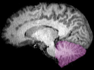
People with A-T begin life with neurological deficits that become devastating through progressive loss of function in a part of the brain called the cerebellum, which leads to severe difficulty with movement and coordination. A-T patients also suffer frequent infections due to their weakened immune systems and have an increased risk for cancer. The disease is caused by lost function in a gene, ATM, that normally repairs damaged DNA in the cells and preserves normal function.
Developing a human neural cell model to understand A-T’s neurodegenerative process — and create a platform for testing new treatments — was critical because the disease presents differently in humans and laboratory animals. Scientists commonly use mouse models to study A-T, but mice with the disease do not experience the more debilitating effects that humans do. In mice with A-T, the cerebellum appears normal and they do not exhibit the obvious degeneration seen in the human brain.
Lee and colleagues used iPS cell–derived neural cells developed from skin cells of A-T patients with a specific type of genetic mutation to create a disease-in-a-dish model. In the laboratory, researchers were able to model the characteristics of A-T, such as the cell’s lack of ATM protein and its inability to repair DNA damage. The model also allowed the researchers to identify potential new therapeutic drugs, called small molecule read-through (SMRT) compounds, that increase ATM protein activity and improve the model cells’ ability to repair damaged DNA.
“A-T patients with no ATM activity have severe disease but patients with some ATM activity do much better,” Lee said. “This makes our discovery promising, because even a small increase in the ATM activity induced by the SMRT drug can potentially translate to positive effects for patients, slowing disease progression and hopefully improving their quality of life.”
These studies suggest that SMRT compounds may have positive effects on all other cell types in the body, potentially improving A-T patients’ immune function and decreasing their susceptibility to cancer.
Additionally, the patient-specific iPS cell–derived neural cells in this study combined with the SMRT compounds can be an invaluable tool for understanding the development and progression of A-T. This iPS cell–neural cell A-T disease model also can be a platform to identify more potent SMRT drugs. The SMRT drugs identified using this model can potentially be applied to most other genetic diseases with the same type of mutations.
Notes about this neurogenetics and ataxia telangiectasia research
This research was supported by training and research grants from the California Institute of Regenerative Medicine, the National Institutes of Health, APRAT, A-T Ease and Scott Richards Foundation.
Written by Shaun Mason
Contact: Shaun Mason – UCLA
Source: University of California at Los Angeles press release
Image Source: The MRI brain scan with the cerebellum highlighted is available in the public domain.
Original Research: Abstract for “SMRT compounds abrogate cellular phenotypes of ataxia telangiectasia in neural derivatives of patient-specific hiPSCs” by Peiyee Lee, Nathan T. Martin, Kotoka Nakamura, Soheila Azghadi, Mandana Amiri, Uri Ben-David, Susan Perlman, Richard A. Gatti, Hailiang Hu and William E. Lowry in Nature Communications. Published online May 7 2013 doi:10.1038/ncomms2824


