

Summary: Functional connectivity in the human brain changes in two distinct ways during adolescence.
Source: University of Cambridge
New brain networks come ‘online’ during adolescence, allowing teenagers to develop more complex adult social skills, but potentially putting them at increased risk of mental illness, according to new research published in the Proceedings of the National Academy of Sciences (PNAS).
Adolescence is a time of major change in life, with increasing social and cognitive skills and independence, but also increased risk of mental illness. While it is clear that these changes in the mind must reflect developmental changes in the brain, it has been unclear how exactly the function of the human brain matures as people grow up from children to young adults.
A team based in the University of Cambridge and University College London has published a major new research study that helps us understand more clearly the development of the adolescent brain.
The study collected functional magnetic resonance imaging (fMRI) data on brain activity from 298 healthy young people, aged 14-25 years, each scanned on one to three occasions about 6 to 12 months apart. In each scanning session, the participants lay quietly in the scanner so that the researchers could analyse the pattern of connections between different brain regions while the brain was in a resting state.
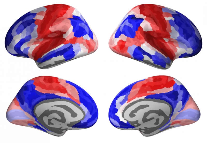
The team discovered that the functional connectivity of the human brain – in other words, how different regions of the brain ‘talk’ to each other – changes in two main ways during adolescence.
The brain regions that are important for vision, movement, and other basic faculties were strongly connected at the age of 14 and became even more strongly connected by the age of 25. This was called a ‘conservative’ pattern of change, as areas of the brain that were rich in connections at the start of adolescence become even richer during the transition to adulthood.
However, the brain regions that are important for more advanced social skills, such as being able to imagine how someone else is thinking or feeling (so-called theory of mind), showed a very different pattern of change. In these regions, connections were redistributed over the course of adolescence: connections that were initially weak became stronger, and connections that were initially strong became weaker. This was called a ‘disruptive’ pattern of change, as areas that were poor in their connections became richer, and areas that were rich became poorer.
By comparing the fMRI results to other data on the brain, the researchers found that the network of regions that showed the disruptive pattern of change during adolescence had high levels of metabolic activity typically associated with active re-modelling of connections between nerve cells.
Dr Petra Vértes, joint senior author of the paper and a Fellow of the mental health research charity MQ, said: “From the results of these brain scans, it appears that the acquisition of new, more adult skills during adolescence depends on the active, disruptive formation of new connections between brain regions, bringing new brain networks ‘online’ for the first time to deliver advanced social and other skills as people grow older.”
Professor Ed Bullmore, joint senior author of the paper and head of the Department of Psychiatry at Cambridge, said: “We know that depression, anxiety and other mental health disorders often occur for the first time in adolescence – but we don’t know why. These results show us that active re-modelling of brain networks is ongoing during the teenage years and deeper understanding of brain development could lead to deeper understanding of the causes of mental illness in young people.”
Measuring functional connectivity in the brain presents particular challenges, as Dr František Váša, who led the study as a Gates Cambridge Trust PhD Scholar, and is now at King’s College London, explained.
“Studying brain functional connectivity with fMRI is tricky as even the slightest head movement can corrupt the data – this is especially problematic when studying adolescent development as younger people find it harder to keep still during the scan,” he said. “Here, we used three different approaches for removing signatures of head movement from the data, and obtained consistent results, which made us confident that our conclusions are not related to head movement, but to developmental changes in the adolescent brain.”
Funding: The study was supported by the Wellcome Trust.
Source:
University of Cambridge
Media Contacts:
Craig Brierley – University of Cambridge
Image Source:
The image is credited to Frantisek Vasa.
Original Research: Open access
“Conservative and disruptive modes of adolescent change in human brain functional connectivity”. Váša, F et al.
PNAS doi:10.1073/pnas.1906144117.
Abstract
Conservative and disruptive modes of adolescent change in human brain functional connectivity
Adolescent changes in human brain function are not entirely understood. Here, we used multiecho functional MRI (fMRI) to measure developmental change in functional connectivity (FC) of resting-state oscillations between pairs of 330 cortical regions and 16 subcortical regions in 298 healthy adolescents scanned 520 times. Participants were aged 14 to 26 y and were scanned on 1 to 3 occasions at least 6 mo apart. We found 2 distinct modes of age-related change in FC: “conservative” and “disruptive.” Conservative development was characteristic of primary cortex, which was strongly connected at 14 y and became even more connected in the period from 14 to 26 y. Disruptive development was characteristic of association cortex and subcortical regions, where connectivity was remodeled: connections that were weak at 14 y became stronger during adolescence, and connections that were strong at 14 y became weaker. These modes of development were quantified using the maturational index (MI), estimated as Spearman’s correlation between edgewise baseline FC (at 14 y, [Math Processing Error]) and adolescent change in FC ([Math Processing Error]), at each region. Disruptive systems (with negative MI) were activated by social cognition and autobiographical memory tasks in prior fMRI data and significantly colocated with prior maps of aerobic glycolysis (AG), AG-related gene expression, postnatal cortical surface expansion, and adolescent shrinkage of cortical thickness. The presence of these 2 modes of development was robust to numerous sensitivity analyses. We conclude that human brain organization is disrupted during adolescence by remodeling of FC between association cortical and subcortical areas.


