

The brain still has a lot to learn about itself. Scientists at the Virginia Tech Carilion Research Institute have made a key finding of the striking differences in how the brain’s cells can change through experience.
Their results were published this week in PLOS ONE.
“Neurons can undergo long-term changes in response to experience such as learning, emotions, or other activity,” said Michael Friedlander, executive director of the Virginia Tech Carilion Research Institute. Friedlander co-authored the paper with his former graduate student and postdoctoral fellow, Ignacio Saez. “Neuroscientists have focused much of their attention on understanding the neuroplasticity of the connections between nerve cells called synapses.”
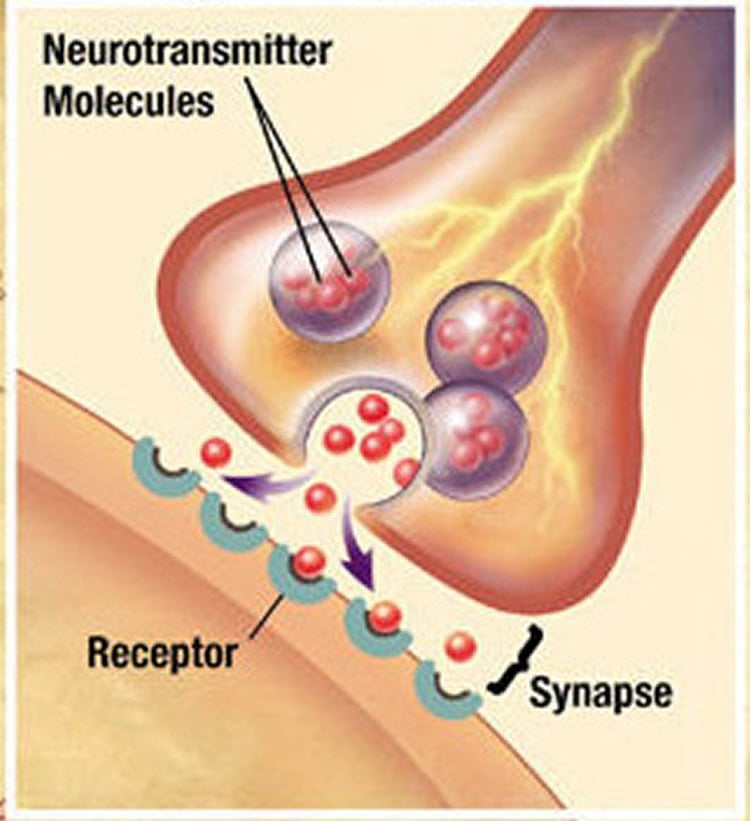
Synapses, the specialized connections between neurons, work by translating an electrical signal from one neuron into a chemical signal to modify the receiving neuron. The chemical signal triggers an electrical signal in the receiving neuron, and the process continues.
Synapses may become stronger or weaker by changing efficiency of the chemical communication process in response to repeated bouts of co-activation of the two interconnected neurons. This process, called synaptic plasticity, can cause changes that persist beyond the co-activation period for mere minutes through a lifetime.
Outside experience can be internalized as a physical reorganization of the brain’s synaptic communication process. This is especially important during the brain’s development but also throughout life as experiences such as learning continually modify the brain’s synaptic circuitry.
“Until recently, scientists had thought that most synapses of a similar type and in a similar location in the brain behaved in a similar fashion with respect to how experience induces plasticity,” Friedlander said. “In our work, however, we found dramatic differences in the plasticity response, even between neighboring synapses in response to identical activity experiences.”
Friedlander and Saez reported that neurons whose excitatory synapses are in a certain states of plasticity, based on previous experiences, assort themselves into groups to converge onto specific individual neurons in the developing brain.
“Individual neurons whose synapses are most likely to strengthen in response to a certain experience are more likely to connect to certain partner neurons, while those whose synapses weaken in response to a similar experience are more likely to connect to other partner neurons,” Friedlander said. “The neurons whose synapses do not change at all in response to that same experience are more likely to connect to yet other partner neurons, forming a more stable but non-plastic network.”
The researchers observed this like-type synaptic plasticity buddy system in a rodent model, using an isolated part of the cerebral cortex responsible for processing vision. The scientists recorded electrical activity from individual neurons after activating groups of neighboring neurons. They then compared that recording to the electrical activity elicited in response to the activation of only a single neighboring neuron. The synapses were trained by repeating the activation process, to mimic learning.
When the scientists applied a pharmacological agent to the neurons that blocked synaptic inhibition, they saw that training elicited more dramatic and varied plasticity at excitatory synapses. The plasticity responses of different groups of synapses on a given neuron were more similar when inhibition was blocked, which effectively grouped together like-type neurons by their learning responses.
“While we’ve known for years that neurons of similar types tend to richly interconnect, this is the first demonstration that such assortment processes apply to synaptic plasticity,” Friedlander said. “Such a result has implications for enhanced learning paradigms, as well as for better understanding the dynamic network properties of the large-scale neuronal networks in the living brain.”
Source: Ashley WennersHerron – Virginia Tech
Image Credit: The image is in the public domain
Original Research: Full open access research for “Role of GABAA-Mediated Inhibition and Functional Assortment of Synapses onto Individual Layer 4 Neurons in Regulating Plasticity Expression in Visual Cortex” by Ignacio Saez, and Michael J. Friedlander in PLOS ONE. Published online February 3 2016 doi:10.1371/journal.pone.0147642
Abstract
Role of GABAA-Mediated Inhibition and Functional Assortment of Synapses onto Individual Layer 4 Neurons in Regulating Plasticity Expression in Visual Cortex
Layer 4 (L4) of primary visual cortex (V1) is the main recipient of thalamocortical fibers from the dorsal lateral geniculate nucleus (LGNd). Thus, it is considered the main entry point of visual information into the neocortex and the first anatomical opportunity for intracortical visual processing before information leaves L4 and reaches supra- and infragranular cortical layers. The strength of monosynaptic connections from individual L4 excitatory cells onto adjacent L4 cells (unitary connections) is highly malleable, demonstrating that the initial stage of intracortical synaptic transmission of thalamocortical information can be altered by previous activity. However, the inhibitory network within L4 of V1 may act as an internal gate for induction of excitatory synaptic plasticity, thus providing either high fidelity throughput to supragranular layers or transmittal of a modified signal subject to recent activity-dependent plasticity. To evaluate this possibility, we compared the induction of synaptic plasticity using classical extracellular stimulation protocols that recruit a combination of excitatory and inhibitory synapses with stimulation of a single excitatory neuron onto a L4 cell. In order to induce plasticity, we paired pre- and postsynaptic activity (with the onset of postsynaptic spiking leading the presynaptic activation by 10ms) using extracellular stimulation (ECS) in acute slices of primary visual cortex and comparing the outcomes with our previously published results in which an identical protocol was used to induce synaptic plasticity between individual pre- and postsynaptic L4 excitatory neurons. Our results indicate that pairing of ECS with spiking in a L4 neuron fails to induce plasticity in L4-L4 connections if synaptic inhibition is intact. However, application of a similar pairing protocol under GABAARs inhibition by bath application of 2μM bicuculline does induce robust synaptic plasticity, long term potentiation (LTP) or long term depression (LTD), similar to our results with pairing of pre- and postsynaptic activation between individual excitatory L4 neurons in which inhibitory connections are not activated. These results are consistent with the well-established observation that inhibition limits the capacity for induction of plasticity at excitatory synapses and that pre- and postsynaptic activation at a fixed time interval can result in a variable range of plasticity outcomes. However, in the current study by virtue of having two sets of experimental data, we have provided a new insight into these processes. By randomly mixing the assorting of individual L4 neurons according to the frequency distribution of the experimentally determined plasticity outcome distribution based on the calculated convergence of multiple individual L4 neurons onto a single postsynaptic L4 neuron, we were able to compare then actual ECS plasticity outcomes to those predicted by randomly mixing individual pairs of neurons. Interestingly, the observed plasticity profiles with ECS cannot account for the random assortment of plasticity behaviors of synaptic connections between individual cell pairs. These results suggest that connections impinging onto a single postsynaptic cell may be grouped according to plasticity states.
“Role of GABAA-Mediated Inhibition and Functional Assortment of Synapses onto Individual Layer 4 Neurons in Regulating Plasticity Expression in Visual Cortex” by Ignacio Saez, and Michael J. Friedlander in PLOS ONE. Published online February 3 2016 doi:10.1371/journal.pone.0147642


