

Summary: Researchers successfully transform stem cells derived from skin cells into cells that behave similarly to the human midbrain.
Source: University of Luxembourg.
The most complex organ in humans is the brain. Due to its complexity and, of course, for ethical reasons, it is extremely difficult to do scientific experiments on it – ones that could help us to understand neurodegenerative diseases like Parkinson’s, for example. Scientists at the Luxembourg Centre for Systems Biomedicine (LCSB) of the University of Luxembourg have now succeeded in turning human stem cells derived from skin samples into tiny, three-dimensional, brain-like cultures that behave very similarly to cells in the human midbrain. In the researchers’ petri dishes, different cell types develop, connect into a network, exchange signals and produce metabolic products typical of the active brain.
“Our cell cultures open new doors to brain research,” says Prof. Dr. Jens Schwamborn, in whose LCSB research group Developmental & Cellular Biology the research work was done. “We can now use them to study the causes of Parkinson’s disease and how it could possibly be effectively treated.” The team publishes its results today in the prestigious scientific journal “Stem Cell Reports“.
The human midbrain is of particular interest to Parkinson’s researchers: it is the seat of the tissue structure known medically as the substantia nigra. Here, nerve cells – specifically dopaminergic neurons – produce the messenger dopamine. Dopamine is needed to maintain smooth body movements. If the dopaminergic neurons die off, then the person affected develops tremors and muscle rigidity, the distinctive symptoms of Parkinson’s disease. For ethical reasons, researchers cannot take cells from the substantia nigra to study them. Research groups around the world are therefore working on cultivating three-dimensional structures of the midbrain in petri dishes. The LCSB team led by stem cell researcher Jens Schwamborn is one such group.
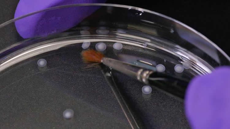
The LCSB scientists worked with so-called induced pluripotent stem cells – stem cells that cannot produce a complete organism, but which can be transformed into all cell types of the human body. The procedures required for converting the stem cells into brain cells were developed by Anna Monzel as part of her doctoral thesis, which she is doing in Schwamborn’s group. “I had to develop a special, precisely defined cocktail of growth factors and a certain treatment method for the stem cells, so that they would differentiate in the desired direction,” Monzel describes her approach. To do this, she was able to draw on extensive preparatory work that had been done in Schwamborn’s team the years before. The pluripotent stem cells in the petri dishes multiplied and spread out into a three-dimensional supporting structure – producing tissue-like cell cultures.
“Our subsequent examination of these artificial tissue samples revealed that various cell types characteristic of the midbrain had developed,” says Jens Schwamborn. “The cells can transmit and process signals. We were even able to detect dopaminergic cells – just like in the midbrain.” This fact makes the LCSB scientists’ results of extraordinary interest to Parkinson’s researchers worldwide, as Schwamborn stresses: “On our new cell cultures, we can study the mechanisms that lead to Parkinson’s much better than was ever the case before. We can test what effects environmental impacts such as pollutants have on the onset of the disease, whether there are new active agents that could possibly relieve the symptoms of Parkinson’s – or whether the disease could even be cured from its very cause. We will be performing such investigations next.”
The development of the brain-like tissue cultures not only opens doors to new research approaches. It can also help to reduce the amount of animal testing in brain research. The cell cultures in the petri dishes are of human origin, and in some aspects resemble human brains more than the brains of lab animals such as rats or mice do. Therefore, the structures of human brains and its modes of function can be modelled in different ways than it is possible in animals. “There are also attractive economic opportunities in our approach,” Jens Schwamborn explains: “The production of tissue cultures is highly elaborate. In the scope of our spin-off Braingineering Technologies Sarl, we will be developing technologies by which we can provide the cultures for a fee to other labs or the pharmaceutical industry for their research.”
Source: Thomas Klein – University of Luxembourg
Image Source: NeuroscienceNews.com image is credited ScienceRelations/University of Luxembour.
Original Research: Full open access research for “Derivation of Human Midbrain-Specific Organoids from Neuroepithelial Stem Cells” by Anna S. Monzel, Lisa M. Smits, Kathrin Hemmer, Siham Hachi, Edinson Lucumi Moreno, Thea van Wuellen, Javier Jarazo, Jonas Walter, Inga Brüggemann, Ibrahim Boussaad, Emanuel Berger, Ronan M.T. Fleming, Silvia Bolognin, and Jens C. Schwamborn in Sten Cell Reports. Published online April 13 2017 doi:10.1016/j.stemcr.2017.03.010
[cbtabs][cbtab title=”MLA”]University of Luxembourg “Brain Tissue From a Petri Dish.” NeuroscienceNews. NeuroscienceNews, 13 April 2017.
<https://neurosciencenews.com/stem-cell-brain-tissue-6407/>.[/cbtab][cbtab title=”APA”]University of Luxembourg (2017, April 13). Brain Tissue From a Petri Dish. NeuroscienceNew. Retrieved April 13, 2017 from https://neurosciencenews.com/stem-cell-brain-tissue-6407/[/cbtab][cbtab title=”Chicago”]University of Luxembourg “Brain Tissue From a Petri Dish.” https://neurosciencenews.com/stem-cell-brain-tissue-6407/ (accessed April 13, 2017).[/cbtab][/cbtabs]
Abstract
Derivation of Human Midbrain-Specific Organoids from Neuroepithelial Stem Cells
Highlights
•Generation of a human in vitro midbrain model from neural precursor cells
•Midbrain organoids show neuronal, astroglial, and oligodendrocyte differentiation
•Midbrain organoids contain spatially organized groups of dopaminergic neurons
•Detection of synaptic connections, electrophysiological activity, and myelination
Summary
Research on human brain development and neurological diseases is limited by the lack of advanced experimental in vitro models that truly recapitulate the complexity of the human brain. Here, we describe a robust human brain organoid system that is highly specific to the midbrain derived from regionally patterned neuroepithelial stem cells. These human midbrain organoids contain spatially organized groups of dopaminergic neurons, which make them an attractive model for the study of Parkinson’s disease. Midbrain organoids are characterized in detail for neuronal, astroglial, and oligodendrocyte differentiation. Furthermore, we show the presence of synaptic connections and electrophysiological activity. The complexity of this model is further highlighted by the myelination of neurites. The present midbrain organoid system has the potential to be used for advanced in vitro disease modeling and therapy development.
“Context-dependent spatially periodic activity in the human entorhinal cortex” by Yu Zhang, Chengzu Long, Hui Li, John R. McAnally, Kedryn K. Baskin, John M. Shelton, Rhonda Bassel-Duby, and Eric N. Olson in PNAS. Published online April 10 2017 doi:10.1073/pnas.1701352114


