

Summary: A new study challenges existing theories about spinal cord neurons. New findings suggest neurological signals originate from a major, scattered network of cells that send signals to only a few other neurons.
Source: University of Copenhagen
With a study of the network between nerve and muscle cells in turtles, researchers from the University of Copenhagen have gained new insight into the way in which movements are generated and maintained. In the long term, the new knowledge may have an impact on the treatment of, for example, ALS and spinal cord injuries.
For most people, it is easy to put one leg in front of the other and keep walking. The ability to do it is second nature or, so to speak, in our bones. Quite literally.
‘Most movements are actually generated in the spinal cord. Naturally, there is a conversation with high-ranking parts of the nervous system, such as the cerebrum, but there are also reflexes that simply stem from the back’, says Associate Professor and Head of Research Rune W. Berg from the Department of Neuroscience at the University of Copenhagen.
He and his research group are behind a study of the network between nerve and muscle cells which has been published in the renowned scientific journal Nature Communications, and which provides completely new insight into the ways in which movements are generated and maintained.
Turtle Crawl
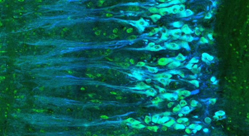
In the study, the research group used electrodes to study the spinal cord reflex of turtles when they scratched themselves with one hind leg. A reflex also found in dogs, cats and a number of other mammals.
Humans are likewise equipped with a variety of spinal reflexes. And although in terms of the evolution, we are rather distant from the turtle, scientists believe that many of the basic mechanisms are the same.
Thus, when the turtle rhythmically scratches itself using crawl movements from its hind leg, the fireworks of lightning quick neurological impulses that are set off inside the shell are not far from the mechanisms that also trigger our own muscles.
From Metronome to Network
So far, it has been a common assumption that the activation of muscle neurons originates from some sort of command centre that sends a signal to many cells at one time.
‘Because the origin of movement has been difficult to find, it has long been assumed that it is a small core that sets the pace. Like some kind of metronome. But our data has shown that it may in fact be a large network’, says Assistant Professor Henrik Lindén from the research group behind the study.
To test whether it was a matter of small command units or a large network, the researchers compared the relatively quiet rhythm of the turtle’s movement with the rapid neurological impulses from the spine.
To the surprise of the research group, the measurements showed no evidence of correlation – and thus no evidence that the neurological signals in multiple cells should have originated from the same source, which would indeed have been the case if it had been a command centre that signalled to multiple cells at the same time.
Instead, the researchers now believe that neurological signals originate from a major, scattered network of cells, each of which sends signals to only a few other cells. A result which the group has subsequently replicated in computer models of a simulated, simple nervous system.
Potential for ALS and Spinal Cord Injuries
With these results, researchers have come a step closer to precisely understanding where and how movements are actually generated.
‘If we do not know enough about the network and how it works, we grope a bit in the dark when it comes to treatment. Conversely, once we gain insight into the principles behind the distribution of the network, and which cell types are important, we can better put the treatment of neurological disorders on the right track’, says Rune W. Berg.
Amongst others, he emphasises neurological disorders such as ALS as well as spinal cord injuries, for example from traffic accidents, as areas where increased knowledge about the spinal nervous system can lead to advances in treatment in the long term.
Likewise, new insights from basic research into the neurons of the spinal cord may benefit other parts of the neurology, for example in connection with cot death, which is associated with defects in brainstem activity.
Now, the next step for the research group is to continue the mapping of the scattered neurological network with optical measurements that allow them to track the activity simultaneously over a larger area.
Source:
University of Copenhagen
Media Contacts:
Rune W. Berg – University of Copenhagen
Image Source:
The image is credited to University of Copenhagen.
Original Research: Open access
“Decoupling of timescales reveals sparse convergent CPG network in the adult spinal cord”. Marija Radosevic, Alex Willumsen, Peter C. Petersen, Henrik Lindén, Mikkel Vestergaard & Rune W. Berg.
Nature Communications doi:10.1038/s41467-019-10822-9.
Abstract
Decoupling of timescales reveals sparse convergent CPG network in the adult spinal cord
During the generation of rhythmic movements, most spinal neurons receive an oscillatory synaptic drive. The neuronal architecture underlying this drive is unknown, and the corresponding network size and sparseness have not yet been addressed. If the input originates from a small central pattern generator (CPG) with dense divergent connectivity, it will induce correlated input to all receiving neurons, while sparse convergent wiring will induce a weak correlation, if any. Here, we use pairwise recordings of spinal neurons to measure synaptic correlations and thus infer the wiring architecture qualitatively. A strong correlation on a slow timescale implies functional relatedness and a common source, which will also cause correlation on fast timescale due to shared synaptic connections. However, we consistently find marginal coupling between slow and fast correlations regardless of neuronal identity. This suggests either sparse convergent connectivity or a CPG network with recurrent inhibition that actively decorrelates common input.


