

Summary: Researchers observe white matter changes in NFL football players who reported concussions.
Source: Johns Hopkins Medicine.
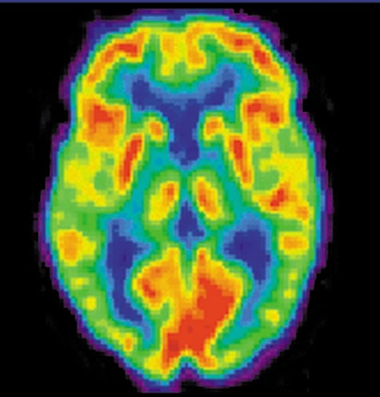
In a small study of young or recently retired NFL players, researchers at Johns Hopkins report finding evidence of brain injury and repair that is visible on imaging from the players compared to a control group of men without a history of concussion.
In a report on the study that used positron emission tomography (PET) and MRI, published in JAMA Neurology on Nov. 28, the researchers highlighted the value of PET imaging to monitor a marker of injury and repair in the brains of NFL players and athletes in other contact sports.
The new research builds on a rising tide of anecdotal evidence and a few scientific studies suggesting that people with repeated concussive head injuries incurred while playing football, hockey or boxing are at higher-than-normal risk of developing the neurodegenerative disease called chronic traumatic encephalopathy (CTE). CTE is associated with memory deficits, confusion, poor decision-making and later onset of dementia.
However, because CTE is often only diagnosed at autopsy, and because similar symptoms may occur in people without repeated head injuries, researchers, including those at Johns Hopkins Medicine, have been developing methods to better visualize tissue damage in the living brain to demonstrate better cause and effect.
“The exciting part of our new findings is that we now believe we have a useful tool to monitor the brains of NFL players and athletes in other contact sports,” says Jennifer Coughlin, M.D., assistant professor of psychiatry and behavioral sciences at Johns Hopkins. “We can measure TSPO, a PET biomarker of brain injury, in these younger players, and we can now begin to follow it over time to see if the brain is repairing itself or not.”
In early 2015, the Johns Hopkins research team published PET imaging results showing higher levels of this same biomarker in the brains of nine elderly former NFL players compared to control participants. However, since they initially studied elderly players who were many years from play, the researchers were unable to tell if the findings were also linked to aging and vascular disease, independent of past NFL play.
For the new study, the researchers collected PET imaging data from 11 men without a history of concussion and compared the scans to those of 12 young NFL players, all of whom were still active or had retired within the past 12 years. All players had a self-reported history of at least one concussion. These players were an average age of 31 years old. About 80 percent were Caucasian and 20 percent were African-American. The control participants were matched to the players by body mass index, age and education level.
The PET imaging was acquired using a radioactive chemical that binds to translocator protein 18 kDa (TSPO), which is normally found at low levels in healthy brain tissue. Since TSPO is increased during cellular response to brain injury, high levels of the TSPO signal on each PET scan can indicate where injury and reparative processes occur.
The researchers found higher radiotracer binding to TSPO in players compared to control participants in eight of the 12 brain regions studied. These regions included the hippocampus, a region functionally involved in memory.
Separately, the researchers examined data from MRI scans to look for structural changes in the brains of the study participants. They found no evidence of brain tissue loss in players compared to control participants in any of the brain regions examined, yet they did find some evidence of white matter changes in the players’ brains.
“We suspect that when the brain moves during a hard hit, it causes a shearing injury of the white matter fibers that travel across the brain,” says Coughlin.
Coughlin cautioned that there are some limitations to the imaging technique. For example, the radiotracer used in the PET scans doesn’t work well in people with a specific variation in the gene that codes for TSPO protein, which occurs in about one in 10 people of European descent. Also, the researchers observed that use of creatine supplements — taken by athletes to improve performance — may interfere with the imaging results, necessitating further study of this effect before including participants taking creatine.
“With further research using this technology, we may better understand the relationship between concussion and brain damage,” says Coughlin. “Further understanding may help inform players of associated risk, and will allow us to test preventive and therapeutic interventions that may improve the lives of players.”
According to Centers for Disease Control and Prevention estimates, anywhere from 1.6 to 3.8 million concussions happen each year in the U.S. because of sports or recreational activities.
Other researchers contributing to the study include Yuchuan Wang, Il Minn, Nicholas Bienko, Emily Ambinder, Xin Xu, Matthew Peters, John Dougherty, Melin Vranesic, Soo Min Koo, Hye-Hyun Ahn, Merton Lee, Chris Cottrell, Haris Sair, Akira Sawa, Cynthia Munro, Robert Dannals, Constantine Lyketsos, Gwenn Smith, Brian Caffo, Susumu Mori and Martin Pomper of The Johns Hopkins University; Christopher Nowinski of Boston University; Michael Kassiou of the University of Sydney; and Tomas Guilarte of Florida International University.
Funding: The study was funded by the Brain and Behavior Research Foundation, the Alexander Wilson Schweizer Fellowship, the National Institute of Environmental Health Sciences (NIEHS-ES007062) and the GE/NFL Head Health Challenge. The funders had no role in designing, conducting or reporting on the study results.
Source: Vanessa McMains – Johns Hopkins Medicine
Image Source: This NeuroscienceNews.com image is credited to NIH.
Original Research: Full open access research for “Imaging of Glial Cell Activation and White Matter Integrity in Brains of Active and Recently Retired National Football League Players” by Jennifer M. Coughlin, MD; Yuchuan Wang, PhD; Il Minn, PhD; Nicholas Bienko, MS; Emily B. Ambinder, MD; Xin Xu, MS; Matthew E. Peters, M1; John W. Dougherty, DO; Melin Vranesic, MD; Soo Min Koo, BS; Hye-Hyun Ahn, PhD; Merton Lee, PhD; Chris Cottrell, BS; Haris I. Sair, MD; Akira Sawa, MD, PhD; Cynthia A. Munro, PhD; Christopher J. Nowinski, AB; Robert F. Dannals, PhD; Constantine G. Lyketsos, MD; Michael Kassiou, PhD; Gwenn Smith, PhD; Brian Caffo, PhD; Susumu Mori, PhD; Tomas R. Guilarte, PhD; and Martin G. Pomper, MD, PhD in JAMA Neurology. Published online November 28 2016 doi:10.1001/jamaneurol.2016.3764
[cbtabs][cbtab title=”MLA”]Johns Hopkins Medicine. “Evidence Of Brain Injury Found In Young NFL Players.” NeuroscienceNews. NeuroscienceNews, 29 November 2016.
<https://neurosciencenews.com/nfl-brain-injury-5621/>.[/cbtab][cbtab title=”APA”]Johns Hopkins Medicine. (2016, November 29). Evidence Of Brain Injury Found In Young NFL Players. NeuroscienceNews. Retrieved November 29, 2016 from https://neurosciencenews.com/nfl-brain-injury-5621/[/cbtab][cbtab title=”Chicago”]Johns Hopkins Medicine. “Evidence Of Brain Injury Found In Young NFL Players.” https://neurosciencenews.com/nfl-brain-injury-5621/ (accessed November 29, 2016).[/cbtab][/cbtabs]
Abstract
Imaging of Glial Cell Activation and White Matter Integrity in Brains of Active and Recently Retired National Football League Players
Importance Microglia, the resident immune cells of the central nervous system, play an important role in the brain’s response to injury and neurodegenerative processes. It has been proposed that prolonged microglial activation occurs after single and repeated traumatic brain injury, possibly through sports-related concussive and subconcussive injuries. Limited in vivo brain imaging studies months to years after individuals experience a single moderate to severe traumatic brain injury suggest widespread persistent microglial activation, but there has been little study of persistent glial cell activity in brains of athletes with sports-related traumatic brain injury.
Objective To measure translocator protein 18 kDa (TSPO), a marker of activated glial cell response, in a cohort of National Football League (NFL) players and control participants, and to report measures of white matter integrity.
Design, Setting, and Participants This cross-sectional, case-control study included young active (n = 4) or former (n = 10) NFL players recruited from across the United States, and 16 age-, sex-, highest educational level–, and body mass index–matched control participants. This study was conducted at an academic research institution in Baltimore, Maryland, from January 29, 2015, to February 18, 2016.
Main Outcomes and Measures Positron emission tomography–based regional measures of TSPO using [11C]DPA-713, diffusion tensor imaging measures of regional white matter integrity, regional volumes on structural magnetic resonance imaging, and neuropsychological performance.
Results The mean (SD) ages of the 14 NFL participants and 16 control participants were 31.3 (6.1) years and 27.6 (4.9) years, respectively. Players reported a mean (SD) of 7.0 (6.4) years (range, 1-21 years) since the last self-reported concussion. Using [11C]DPA-713 positron emission tomographic data from 12 active or former NFL players and 11 matched control participants, the NFL players showed higher total distribution volume in 8 of the 12 brain regions examined (P < .004). We also observed limited change in white matter fractional anisotropy and mean diffusivity in 13 players compared with 15 control participants. In contrast, these young players did not differ from control participants in regional brain volumes or in neuropsychological performance.
Conclusions and Relevance The results suggest that localized brain injury and repair, indicated by higher TSPO signal and white matter changes, may be associated with NFL play. Further study is needed to confirm these findings and to determine whether TSPO signal and white matter changes in young NFL athletes are related to later onset of neuropsychiatric symptoms.
“Imaging of Glial Cell Activation and White Matter Integrity in Brains of Active and Recently Retired National Football League Players” by Jennifer M. Coughlin, MD; Yuchuan Wang, PhD; Il Minn, PhD; Nicholas Bienko, MS; Emily B. Ambinder, MD; Xin Xu, MS; Matthew E. Peters, M1; John W. Dougherty, DO; Melin Vranesic, MD; Soo Min Koo, BS; Hye-Hyun Ahn, PhD; Merton Lee, PhD; Chris Cottrell, BS; Haris I. Sair, MD; Akira Sawa, MD, PhD; Cynthia A. Munro, PhD; Christopher J. Nowinski, AB; Robert F. Dannals, PhD; Constantine G. Lyketsos, MD; Michael Kassiou, PhD; Gwenn Smith, PhD; Brian Caffo, PhD; Susumu Mori, PhD; Tomas R. Guilarte, PhD; and Martin G. Pomper, MD, PhD in JAMA Neurology. Published online November 28 2016 doi:10.1001/jamaneurol.2016.3764


