Summary: A new study identifies a sub region of the brain that works to form a particular kind of memory: fear-associated with a specific environmental cue or “contextual fear memory.”
Source: Scripps Research Institute.
While the romantic poets’ idea of memories being akin to spirits may have poetic merit, the scientists’ perspective is that memories are concrete, physical entities that can be visualized within various regions of the brain.
Scientists from the Florida campus of The Scripps Research Institute (TSRI) have now for the first time identified a sub-region in the brain that works to form a particular kind of memory: fear-associated with a specific environmental cue or “contextual fear memory.”
The study, recently published in the journal Biological Psychiatry Cognitive Neuroscience and Neuroimaging, was led by TSRI Associate Professor Sathyanarayanan V. Puthanveettil.
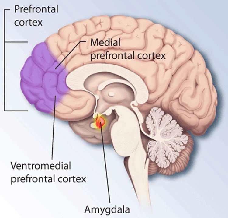
“Much is still unknown about the identities of proteins synthesized to produce long-term memory,” Puthanveettil said. “The most striking observation from the new study is that the medial prefrontal cortex is the site of this early protein synthesis. We have also identified what proteins are newly synthesized in the medial prefrontal cortex.”
In particular, the study showed new protein synthesis in a specific sub-region of the prefrontal cortex known in rodents as the prelimbic. In humans, this area corresponds to the anterior cortex, which has been linked to processing emotional responses.
Initially, Puthanveettil and his colleagues ignored the medial prefrontal cortex because no one believed that it had anything to do with early encoding of long term memories.
However, when they closely examined the effects on the brain of conditioning rodents with a mild foot shock, the scientists found several messenger RNAs recruited to polyribosomes in the medial prefrontal cortex—a clear indication of new protein synthesis there.
Puthanveettil and his colleagues also discovered that if they inhibited new protein synthesis in the prelimbic region right after fear conditioning took place, those memories did not form. But if the researchers waited just a few hours, inhibiting protein synthesis in prelimbic cortex had no impact and the memories took hold. There is temporal and spatial regulation of new protein synthesis in the medial prefrontal cortex.
“It may be that the first wave of protein synthesis is critical for encoding contextual fear memory, while second wave in other sub-regions is important for memory storage,” he said.
It remains to be determined if other sub-regions of the cortex are also be involved in the synthesis of memory proteins.
“The medial prefrontal cortex has many sub-regions,” said TSRI Senior Research Associate Bindu L. Raveendra, co-first author of the study with Valerio Rizzo, Khalid Touzani and Supriya Swarnkar, all of TSRI at the time of the study. “But the specific roles of these sub-regions in encoding, expression and retrieval, as well as their underlying molecular mechanisms, remain to be unraveled.”
Other authors of the study, “Encoding of Contextual Fear Memory Requires De Novo Proteins in The Prelimbic Cortex,” include Beena M. Kadakkuzha and Xin-An Liu of TSRI; Joan Lora and Robert W. Stackman of Florida Atlantic University; and Chao Zhang and Doron Betel of Weill Cornell Medical College.
Funding: The study was supported by the Whitehall Foundation, the National Institutes of Health (grant number 1R21MH096258) and the State of Florida.
Source: Scripps Research Institute
Image Source: This NeuroscienceNews.com image is in the public domain.
Original Research: Abstract for “Encoding of Contextual Fear Memory Requires De Novo Proteins in The Prelimbic Cortex” by Valerio Rizzo, Khalid Touzani, Bindu L. Raveendra, Supriya Swarnkar, Joan Lora, Beena M. Kadakkuzha, Xin-An Liu, Chao Zhang, Doron Betel, Robert W. Stackman, and Sathyanarayanan V. Puthanveettil in Biological Psychiatry Cognitive Neuroscience and Neuroimaging. Published online October 21 2016 doi:10.1016/j.bpsc.2016.10.002
[cbtabs][cbtab title=”MLA”]Scripps Research Institute. “What Does It Take to Make a Memory? Study Says New Proteins.” NeuroscienceNews. NeuroscienceNews, 10 November 2016.
<https://neurosciencenews.com/new-protein-memory-5473/>.[/cbtab][cbtab title=”APA”]Scripps Research Institute. (2016, November 10). What Does It Take to Make a Memory? Study Says New Proteins. NeuroscienceNews. Retrieved November 10, 2016 from https://neurosciencenews.com/new-protein-memory-5473/[/cbtab][cbtab title=”Chicago”]Scripps Research Institute. “What Does It Take to Make a Memory? Study Says New Proteins.” https://neurosciencenews.com/new-protein-memory-5473/ (accessed November 10, 2016).[/cbtab][/cbtabs]
Abstract
Encoding of Contextual Fear Memory Requires De Novo Proteins in The Prelimbic Cortex
Background
Despite our understanding of the significance of the prefrontal cortex in the consolidation of long-term memories (LTM), its role in the encoding of LTM remains elusive. Here we investigated the role of new protein synthesis in the mouse medial prefrontal cortex (mPFC) in encoding contextual fear memory.
Methods
Because a change in the association of mRNAs to polyribosomes is an indicator of new protein synthesis, we assessed the changes in polyribosome-associated mRNAs in the mPFC following contextual fear conditioning (CFC) in the mouse. Differential gene expression in mPFC was identified by polyribosome profiling (n = 18). The role of new protein synthesis in mPFC was determined by focal inhibition of protein synthesis (n = 131) and by intra-prelimbic cortex manipulation (n = 56) of Homer 3, a candidate identified from polyribosome profiling.
Results
We identified several mRNAs that are differentially and temporally recruited to polyribosomes in the mPFC following CFC. Inhibition of protein synthesis in the prelimbic (PL), but not in the anterior cingulate cortex (ACC) region of the mPFC immediately after CFC disrupted encoding of contextual fear memory. Intriguingly, inhibition of new protein synthesis in the PL 6 hours after CFC did not impair encoding. Furthermore, expression of Homer 3, an mRNA enriched in polyribosomes following CFC, in the PL constrained encoding of contextual fear memory.
Conclusions
Our studies identify several molecular substrates of new protein synthesis in the mPFC and establish that encoding of contextual fear memories require new protein synthesis in PL subregion of mPFC.
“Encoding of Contextual Fear Memory Requires De Novo Proteins in The Prelimbic Cortex” by Valerio Rizzo, Khalid Touzani, Bindu L. Raveendra, Supriya Swarnkar, Joan Lora, Beena M. Kadakkuzha, Xin-An Liu, Chao Zhang, Doron Betel, Robert W. Stackman, and Sathyanarayanan V. Puthanveettil in Biological Psychiatry Cognitive Neuroscience and Neuroimaging. Published online October 21 2016 doi:10.1016/j.bpsc.2016.10.002