Summary: Researchers have been able to capture autophagy in super resolution.
Source: Babraham Institute.
Dissecting the cell’s recycling process in super resolution.
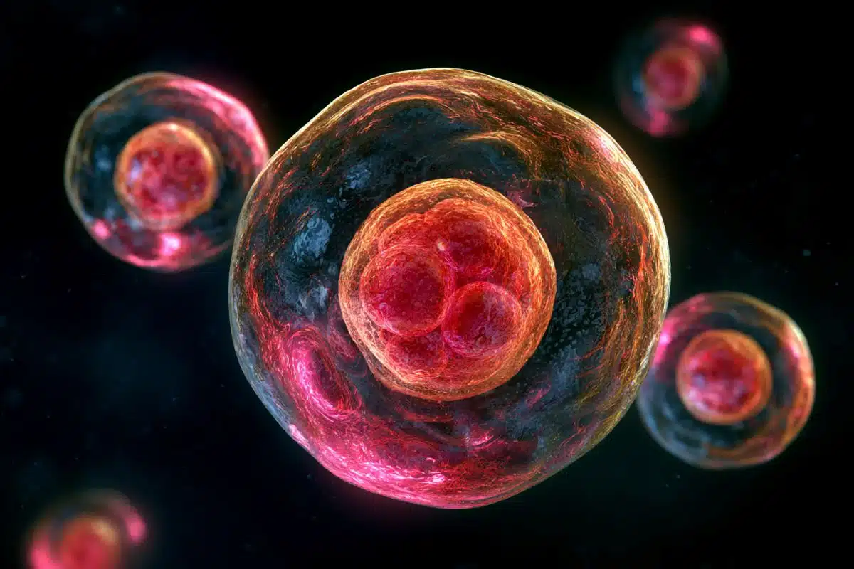
We don’t tend to wrap our recycling waste in bubble wrap but that’s essentially what cells do during the cellular recycling process called autophagy. Using the live imaging capabilities at the Babraham Institute, Institute researchers and their collaborators at Carl Zeiss Microscopy, Munich, and the Francis Crick Institute, London, have viewed the earliest stages of this encapsulation and recycling process in super resolution to reveal what’s happening in unprecedented molecular detail. Their research is published today in the journal Nature Communications.
Derived from the Greek and meaning ‘self-eating’, autophagy describes a process whereby cellular contents are collected and recycled into new molecules and cellular structures; a process of reclaiming the unwanted or damaged and using them to create something useful for the cell. Autophagy is fundamental to the function of our bodies. As the clean-up mechanism for cellular debris, loss of efficiency or glitches in this process are associated with ageing and ageing-related diseases such as Alzheimer’s, rheumatoid arthritis and cancer.
The researchers focused on determining the origin and formation of a structure only seen at the very start of the autophagy process but which gives rise to the main structure (autophagosome; the cellular ‘bubble wrap’) that envelops the content targeted for degradation. Due to its short-lived nature, this transient structure was difficult to characterise. The researchers jointly developed a new comprehensive imaging-based approach for observing autophagy-related structures. At the Babraham Institute this was achieved using live imaging followed by dStorm (direct Stochastic Optical Reconstruction Microscopy). At the Francis Crick Institute in London and the Zeiss Microscopy Labs in Munich, the researchers used a method called FIB-SEM (Focused Ion Beam Scanning Electron Microscopy). By combining the information gathered from these two methods, the researchers were able to identify how the first autophagy structure forms and clarify the protein and membrane associations leading to its development into a fully-fledged autophagosome.

Dr Nicholas Ktistakis, group leader in the Signalling research programme at the Babraham Institute and lead senior author, said: “By combining live imaging with cutting-edge super resolution microscopy techniques, we have been able to characterise the site of autophagy initiation and observe the physical and functional interactions between the proteins involved in autophagy. This has uncovered a new level of detail of the earliest stages of autophagy and provides a general protocol for this type of analysis in other areas of cell biology.
“Knowing more about this process increases our ability to find ways to manipulate or boost it for future therapeutic benefit.”
Funding: This work was supported by the Biotechnology and Biological Sciences Research Council.
Source: Louisa Wood – Babraham Institute
Image Source: This NeuroscienceNews.com image is credited to The Babraham Institute.
Original Research: Full open access research for “Autophagy initiation by ULK complex assembly on ER tubulovesicular regions marked by ATG9 vesicles” by Eleftherios Karanasios, Simon A. Walker, Hanneke Okkenhaug, Maria Manifava, Eric Hummel, Hans Zimmermann, Qashif Ahmed, Marie-Charlotte Domart, Lucy Collinson and Nicholas T. Ktistakis in Nature Communications. Published online August 11 2016 doi:10.1038/ncomms12420
[cbtabs][cbtab title=”MLA”]Babraham Institute. “Putting Autophagy Under the Microscope.” NeuroscienceNews. NeuroscienceNews, 11 August 2016.
<https://neurosciencenews.com/neuroscience-autophagy-4828/>.[/cbtab][cbtab title=”APA”]Babraham Institute. (2016, August 11). Putting Autophagy Under the Microscope. NeuroscienceNews. Retrieved August 11, 2016 from https://neurosciencenews.com/neuroscience-autophagy-4828/[/cbtab][cbtab title=”Chicago”]Babraham Institute. “Putting Autophagy Under the Microscope.” https://neurosciencenews.com/neuroscience-autophagy-4828/ (accessed August 11, 2016).[/cbtab][/cbtabs]
Abstract
Autophagy initiation by ULK complex assembly on ER tubulovesicular regions marked by ATG9 vesicles
Autophagosome formation requires sequential translocation of autophagy-specific proteins to membranes enriched in PI3P and connected to the ER. Preceding this, the earliest autophagy-specific structure forming de novo is a small punctum of the ULK1 complex. The provenance of this structure and its mode of formation are unknown. We show that the ULK1 structure emerges from regions, where ATG9 vesicles align with the ER and its formation requires ER exit and coatomer function. Super-resolution microscopy reveals that the ULK1 compartment consists of regularly assembled punctate elements that cluster in progressively larger spherical structures and associates uniquely with the early autophagy machinery. Correlative electron microscopy after live imaging shows tubulovesicular membranes present at the locus of this structure. We propose that the nucleation of autophagosomes occurs in regions, where the ULK1 complex coalesces with ER and the ATG9 compartment.
“Autophagy initiation by ULK complex assembly on ER tubulovesicular regions marked by ATG9 vesicles” by Eleftherios Karanasios, Simon A. Walker, Hanneke Okkenhaug, Maria Manifava, Eric Hummel, Hans Zimmermann, Qashif Ahmed, Marie-Charlotte Domart, Lucy Collinson and Nicholas T. Ktistakis in Nature Communications. Published online August 11 2016 doi:10.1038/ncomms12420