

Summary: A new study reports brain connectivity appears to be dictated by the spatial architecture of neurons, rather than the cell type-specific cues.
Source: UC Irvine.
A University of California, Irvine-led study reveals that connectivity within the brain appears to be largely dictated by spatial architecture rather than cell type-specific cues. The study was published this month in Cell Reports.
To understand how the brain works, current neuroscience research has focused on mapping the connectome to determine how the brain is built and how it is wired. Much research has been done to map the outputs of defined neuronal populations and to identify what controls them. Researchers in this study used a viral-assisted method to map the direct inputs onto targeted neuronal populations in the brain to compare the inputs to intermingled cell populations with differing behavioral functions.
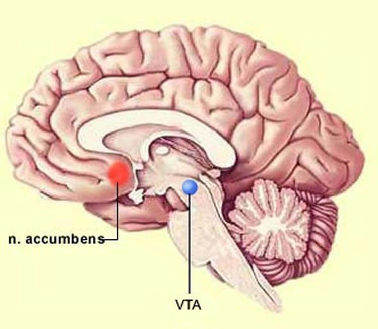
Led by Kevin Beier, PhD, an assistant professor in the Department of Physiology & Biophysics at the UCI School of Medicine, the research team mapped inputs to ventral tegmental area dopamine (VTA) neurons and conducted a rigorous analysis of neuronal connectivity to this region. The cells in the VTA are responsible for a variety of both normal behaviors, such as reward and aversion, as well as pathological behaviors, such as drug addiction and depression. Building on Beier’s previous work published in 2015, the team constructed a comprehensive, high-resolution input-output map of the VTA by standardizing viral reagents and tracing inputs to unique VTA cell types, defined both by the neurotransmitter they released and specific projection targets.
“We used a viral-genetic method to identify the factors that influence connectivity onto different cell populations in the VTA,” said Beier. “In contrast to most studies suggesting specific connectivity patterns, we found that all of the VTA populations that we examined had highly similar inputs. Furthermore, the neurotransmitter released by the neurons we examined – dopamine, GABA, or glutamate – appeared to have little influence on input patterns.”
As the researchers found little evidence for cell type-specific connections, their findings supported a more random connectivity logic, where inputs connect semi-randomly in regions to which the neurons project.
“Interestingly, we found that publicly-available output maps could be used to ‘predict’ our findings. Just knowing where axons from input neurons projected was enough to predict differences in connectivity to different cell types,” Beier explained. “Our findings suggest that researchers may be able to generate global ‘pseudo input maps,’ which previously took years to generate and analyze, simply by using publicly-available resources that can be mined within minutes.”
Recent studies have shown that activation of unique VTA neuronal populations have different behavioral outcomes. Follow-up studies have attempted to identify how control of these populations differs, and how this may be explained by differences in input connectivity. Though this study suggests that differences in anatomical connectivity may be overstated, further testing is needed to determine how anatomical maps relate to function and if this principle applies to brain regions other than the VTA.
Funding: National Institutes of Health, Howard Hughes Medical Institute funded this study.
Source: Anne Warde – UC Irvine
Publisher: Organized by NeuroscienceNews.com.
Image Source: NeuroscienceNews.com image is for illustrative purposes only.
Original Research: Open access research for “Topological Organization of Ventral Tegmental Area Connectivity Revealed by Viral-Genetic Dissection of Input-Output Relations” by Kevin T. Beier, Xiaojing J. Gao, Stanley Xie, Katherine E. DeLoach, Robert C. Malenka, and Liqun Luo in Cell Reports. Published January 2 2019.
doi:10.1016/j.celrep.2018.12.040
[cbtabs][cbtab title=”MLA”]UC Irvine”When It Comes to Brain Connectivity, Cell Location Matters Most.” NeuroscienceNews. NeuroscienceNews, 25 January 2019.
<https://neurosciencenews.com/neuron-location-connectivity-10644/>.[/cbtab][cbtab title=”APA”]UC Irvine(2019, January 25). When It Comes to Brain Connectivity, Cell Location Matters Most. NeuroscienceNews. Retrieved January 25, 2019 from https://neurosciencenews.com/neuron-location-connectivity-10644/[/cbtab][cbtab title=”Chicago”]UC Irvine”When It Comes to Brain Connectivity, Cell Location Matters Most.” https://neurosciencenews.com/neuron-location-connectivity-10644/ (accessed January 25, 2019).[/cbtab][/cbtabs]
Abstract
A Twin Study on the Association Between Psychotic Experiences and Tobacco Use During Adolescence
Viral-genetic tracing techniques have enabled mesoscale mapping of neuronal connectivity by teasing apart inputs to defined neuronal populations in regions with heterogeneous cell types. We previously observed input biases to output-defined ventral tegmental area dopamine (VTA-DA) neurons. Here, we further dissect connectivity in the VTA by defining input-output relations of neurochemically and output-defined neuronal populations. By expanding our analysis to include input patterns to subtypes of excitatory (vGluT2-expressing) or inhibitory (GAD2-expressing) populations, we find that the output site, rather than neurochemical phenotype, correlates with whole-brain inputs of each subpopulation. Lastly, we find that biases in input maps to different VTA neurons can be generated using publicly available whole-brain output mapping datasets. Our comprehensive dataset and detailed spatial analysis suggest that connection specificity in the VTA is largely a function of the spatial location of the cells within the VTA.


