

Researchers at King’s College London have discovered how a molecular ‘scaffold’ which allows key parts of cells to interact, comes apart in dementia and motor neuron disease, revealing a potential new target for drug discovery.
The study, published today in Nature Communications, was funded by the UK Medical Research Council, Wellcome Trust, Alzheimer’s Research UK and the Motor Neurone Disease Association.
Researchers looked at two components of cells: mitochondria, the cell ‘power houses’ which produce energy for the cell;and the endoplasmic reticulum (ER) which makes proteins and stores calcium for signalling processes in the cell. ER and mitochondria form close associations and these interactions enable a number of important cell functions. However the mechanism by which ER and mitochondria become linked has not, until now, been fully understood.
Professor Chris Miller, from the Department of Neuroscience at the Institute of Psychiatry at King’s and lead author of the paper, says: “At the molecular level, many processes go wrong in dementia and motor neuron disease,and one of the puzzles we’re faced with is whether there is a common pathway connecting these different processes. Our study suggests that the loosening of this ‘scaffold’ between the mitochondria and ER in the cell may be a key process in neurodegenerative diseases such as dementia or motor neuron disease.”
By studying cells in a dish, the researchers discovered that an ER protein called VAPB binds to a mitochondrial protein called PTPIP51, to form a ‘scaffold’ enabling ER and mitochondria to form close associations. In fact, by increasing the levels of VAPB and PTPIP51, mitochondria and ER re-organised themselves to form tighter bonds.
Many of the cell’s functions that are controlled by ER-mitochondria associations are disrupted in neurodegenerative diseases, so the researchers studied how the strength of this ‘scaffold’ was affected in these diseases. TDP-43 is a protein which is strongly linked to Amyotrophic Lateral Sclerosis (ALS, a form of motor neuron disease) and Fronto-Temporal Dementia (FTD, the second most common form of dementia), but exactly how the protein causes neurodegeneration is not properly understood. The researchers studied how TDP-43 affected mouse cells in a dish. They found that higher levels of TDP-43 resulted in a loosening of the scaffold which reduced ER-mitochondria bonds,affecting some important cellular functions that are linked to ALS and FTD.
Professor Miller concludes: “Our findings are important in terms of advancing our understanding of basic biology, but may also provide a potential new target for developing new treatments for these devastating disorders.”
Contact: Seil Collins – King’s College London
Source: King’s College London press release
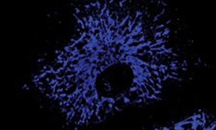
Image Source: The image is adapted from the King’s College London press release
Original Research: Full open access research for “ER–mitochondria associations are regulated by the VAPB–PTPIP51 interaction and are disrupted by ALS/FTD-associated TDP-43” by Radu Stoica, Kurt J. De Vos, Sébastien Paillusson, Sarah Mueller, Rosa M. Sancho, Kwok-Fai Lau, Gema Vizcay-Barrena, Wen-Lang Lin, Ya-Fei Xu, Jada Lewis, Dennis W. Dickson, Leonard Petrucelli, Jacqueline C. Mitchell, Christopher E. Shaw and Christopher C. J. Miller in Nature Communications. Published online June 3 2014 doi:10.1038/ncomms4996
ER–mitochondria associations are regulated by the VAPB–PTPIP51 interaction and are disrupted by ALS/FTD-associated TDP-43
Mitochondria and the endoplasmic reticulum (ER) form tight structural associations and these facilitate a number of cellular functions. However, the mechanisms by which regions of the ER become tethered to mitochondria are not properly known. Understanding these mechanisms is not just important for comprehending fundamental physiological processes but also for understanding pathogenic processes in some disease states. In particular, disruption to ER–mitochondria associations is linked to some neurodegenerative diseases. Here we show that the ER-resident protein VAPB interacts with the mitochondrial protein tyrosine phosphatase-interacting protein-51 (PTPIP51) to regulate ER–mitochondria associations. Moreover, we demonstrate that TDP-43, a protein pathologically linked to amyotrophic lateral sclerosis and fronto-temporal dementia perturbs ER–mitochondria interactions and that this is associated with disruption to the VAPB–PTPIP51 interaction and cellular Ca2+ homeostasis. Finally, we show that overexpression of TDP-43 leads to activation of glycogen synthase kinase-3β (GSK-3β) and that GSK-3β regulates the VAPB–PTPIP51 interaction. Our results describe a new pathogenic mechanism for TDP-43.
“ER–mitochondria associations are regulated by the VAPB–PTPIP51 interaction and are disrupted by ALS/FTD-associated TDP-43” by Radu Stoica, Kurt J. De Vos, Sébastien Paillusson, Sarah Mueller, Rosa M. Sancho, Kwok-Fai Lau, Gema Vizcay-Barrena, Wen-Lang Lin, Ya-Fei Xu, Jada Lewis, Dennis W. Dickson, Leonard Petrucelli, Jacqueline C. Mitchell, Christopher E. Shaw and Christopher C. J. Miller in Nature Communications, June 3 2014 doi:10.1038/ncomms4996


