Summary: Researchers have observed, in real time, the formation and repair of myelin sheaths around nerve fibers.
Source: TUM.
Nerve fibers are surrounded by a myelin sheath. Scientists at the Technical University of Munich (TUM) have now made the first-ever “live” observations of how this protective layer is formed. The team discovered that the characteristic patterns of the myelin layer are determined at an early stage. However, these patterns can be adjusted as needed in a process apparently controlled by the nerve cells themselves.
The myelin sheath surrounding the axons, or nerve fibers, can be compared to the insulation covering an electric wire. Without it, the rapid propagation of electric signals would not be possible. Damage to this insulating layer, for example from illnesses such as multiple sclerosis, may result in serious impairments.
MYELIN SEGMENTS DETERMINE THE TRANSMISSION SPEED
But myelin does not form a continuous coating along the axon. Instead, it is divided into segments. These can vary in length and are separated by gaps known as nodes of Ranvier. For the complex network of the central nervous system to function properly, the speed of the connections is not the only consideration. The crucial factor is the fine tuning: the signals have to reach the right place at exactly the right time. The transmission speed of information through an axon is partly determined by the number and length of the segments.
PATTERNS REMAIN STABLE
The bodies of humans and animals have the ability – at least to some extent – to repair damaged myelin sheaths. Dr. Tim Czopka, a neuroscientist at TUM, has succeeded in making the first-ever “live” observations of this process. Czopka and his team used newly developed markers to visualize the formation myelin segments surrounding axons in the spinal cord of zebrafish.
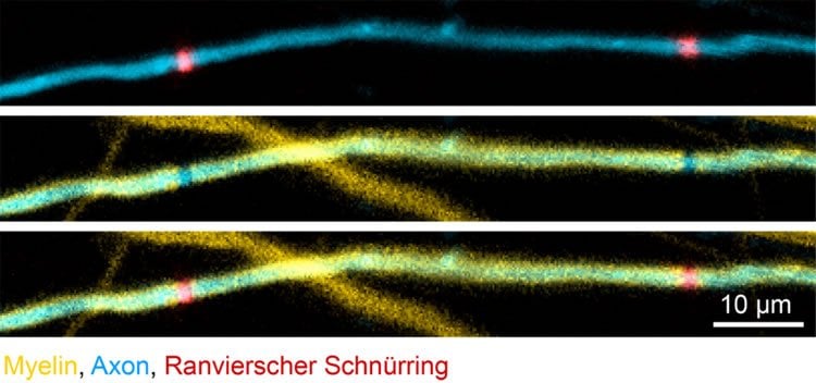
They concluded: Characteristic patterns made up of myelin segments with different lengths along an axon are defined within a few days after myelin formation begins. Although the segments continue growing from that time onward – as the body of the zebrafish grows – the pattern remains preserved.
Following this observation, the researchers destroyed selected segments. “What happened next surprised us,” says Tim Czopka. “After the destruction of the segments, the myelin sheaths began to dynamically remodel. In the end, the damage was repaired and in most cases the original pattern was restored.” The regeneration followed a fixed sequence: First, the adjacent segments expanded to close the gap. A new segment then formed between them, and they contracted to their original size.
AXONS INFLUENCE THE FORMATION OF SEGMENTS
This raises an important question: Who controls the emergence and the restoration of the segment pattern? “Our observations suggest that it is not the oligodendrocytes – the cells that form myelin – that decide where it is formed, but rather the axons,” says Tim Czopka. “You could say that they know best which pattern is needed for the signals to be transmitted at optimal speed.”
The team is now studying how the segment patterns are affected by the targeted stimulation of nerve cell activity and by the neurotransmitters released as a result. “If we can understand the role of the axons in myelin generation and remodelling, it may yield new approaches to controlling it,” explains Czopka. “That would be relevant for the treatment of illnesses such as multiple sclerosis.”
Source: Tim Czopka – TUM
Publisher: Organized by NeuroscienceNews.com.
Image Source: NeuroscienceNews.com image is credited to Czopka / TUM.
Video Source: Video credited to
TUMuenchen1.
Original Research: Open access research in Current Biology.
doi:10.1016/j.cub.2018.01.017
[cbtabs][cbtab title=”MLA”]TUM “Watching Myelin Patterns Form.” NeuroscienceNews. NeuroscienceNews, 14 February 2018.
<https://neurosciencenews.com/myelin-form-8492/>.[/cbtab][cbtab title=”APA”]TUM (2018, February 14). Watching Myelin Patterns Form. NeuroscienceNews. Retrieved February 14, 2018 from https://neurosciencenews.com/myelin-form-8492/[/cbtab][cbtab title=”Chicago”]TUM “Watching Myelin Patterns Form.” https://neurosciencenews.com/myelin-form-8492/ (accessed February 14, 2018).[/cbtab][/cbtabs]
Abstract
Evidence for Myelin Sheath Remodeling in the CNS Revealed by In Vivo Imaging
Highlights
•Differences in myelin sheath length are established in a few days after initiation
•Mature myelin sheaths continue extending to compensate for overall body growth
•Axon myelination patterns remain stable for long periods of time
•Myelin sheath ablation induces remodeling to restore the original myelin pattern
Summary
The length of myelin sheaths affects conduction speed along axons and information propagation. It has recently become clear that myelin may be adaptively modified to modulate circuit function, implying that length remodeling of myelin sheaths should occur. However, direct evidence for such events is lacking. We have investigated how myelination patterns are formed, maintained, and remodeled using long-term imaging and myelin ablation in zebrafish. We demonstrate that length differences between myelin sheaths are established by rapid and variable growth within 3 days after their formation, independently of their time of formation, and even along discontinuously myelinated axons. Afterward, sheaths continue extending at similar rates to compensate for overall animal growth. In consequence, once axon myelination patterns are established, they are maintained over long periods of time. We tested whether mature myelin sheaths can remodel by removing individual sheaths from single axons by targeted ablation. Remarkably, extensive changes in sheath length and number occurred, which frequently restored the original myelination pattern. Our results show that axons can control myelin growth and remodeling, and we provide evidence for a homeostatic control of axon myelination patterns by maintenance and remodeling of myelin sheath length, with implications for circuit development, function, and repair.