When you’re taking a walk around the block, your body is mostly on autopilot—you don’t have to consciously think about alternating which leg you step with or which muscles it takes to lift a foot and put it back down. That’s thanks to a set of cells in your spinal cord that help translate messages between your brain and your motor neurons, which control muscles.
Now, for the first time, researchers have created a method to watch—in real time—the activity of those motor neurons. The new technology, developed by Salk scientists and published in Neuron, is helping researchers understand how spinal cord cells make connections with motor neurons, and how clinicians might be able to repair those connections in patients with spinal cord injuries or neurodegenerative diseases like amyotrophic lateral sclerosis (ALS).
“Using optical methods to be able to watch neuron activity has been a dream over the past decade,” says Samuel Pfaff, a professor in Salk’s Gene Expression Laboratory. “Now, it’s one of those rare times when the technology is actually coming together to show you things you hadn’t been able to see before.”
In the past, to measure the activity of neurons—whether in the brain or extending throughout the body—scientists relied on electrodes that could detect the change in electrical voltage inside a cell when it’s activated. But it is tricky to use electrodes to simultaneously record the activity of many different neuron types at once to study how their activity is synchronized.
To get around this shortcoming of electrode readings, Pfaff’s team used a fluorescent sensor protein called GCaMP6f that lights up whenever a neuron is activated. Unlike the electrodes, the protein could easily be added to many different cells at once. When Pfaff and his colleagues added GCaMP6f to motor neurons, they could watch with a microscope which cells were activated in a mouse spinal cord when chemicals that turn on walking circuits were added.
“You don’t need to do any kind of post-image processing to interpret this,” says Pfaff. “These are just raw signals you can see through the eyepiece of a microscope. It’s really a jaw-dropping kind of visualization for a neuroscientist.”

Pfaff’s group used the new method to answer a long-standing question about how a collection of cells in the spinal cord, called the locomotor central pattern generator (CPG), connects to the right motor neurons to allow movements like walking. The CPG, Pfaff says, is where relatively simple signals from the brain—to walk forward, or move your hand off a hot stove—are translated into more complex instructions for motor neurons to control muscles.
“Our nervous system has to make decisions and computations to tell different muscles to contract, or when not to contract, or the amount of force and speed to use when contracting,” Pfaff explains. It’s the CPG that helps make many of these computations, scientists believe. So normal movement requires that CPG neurons in the spinal cord connect to and control when motor neurons fire. But, until now, researchers didn’t know exactly how the CPG cells forged these connections.
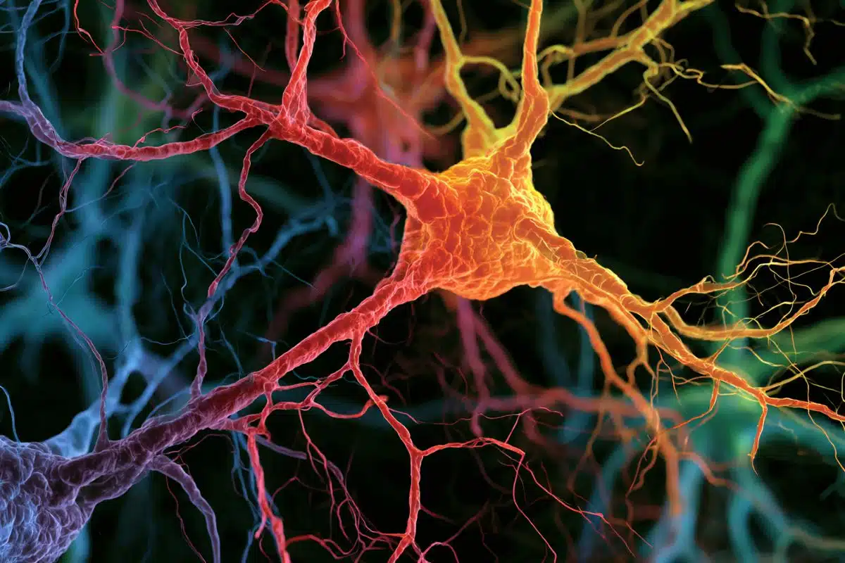
Two-photon imaging let researchers see how different types of motor neurons in the spinal cord alternate activity (shown by bright green fluorescence here) in a cyclical pattern.
By tweaking the locations and identities of motor neurons, and then watching the resulting patterns of activation using their new fluorescent technique, Chris Hinckley in the Pfaff laboratory found that the CPG didn’t rely solely on the cells’ locations to connect to them. Instead, the genetic identity of each subtype of cells—what makes those that control the quadriceps muscle different from those that control the calf muscle for instance—is also important.
That’s a key finding, Pfaff says, for research on how to treat spinal cord injuries and ALS. Currently, many scientists are attempting to turn stem cells into motor neurons, which they then implant into the spinal cord to regenerate damaged connections. Pfaff’s new results, though, suggest that general motor neurons might not do the trick—the best treatment may require the right subtypes of motor neurons. More work, however, is needed to understand the implications of this and exactly how it might translate to disease treatment.
Other researchers on the study were Joseph D. Dekker and Haley O. Tucker of The University of Texas at Austin; and Christopher A. Hinckley, William A. Alaynick, Benjamin W. Gallarda, Marito Hayashi, Kathryn L. Hilde, Shawn P. Driscoll and Tatyana O. Sharpee of the Salk Institute.
Funding: The work and the researchers involved were supported by grants from the National Institutes of Health, Howard Hughes Medical Institute, the Christopher and Dana Reeve Foundation, the Sol Goldman Trust and the Marshall Heritage Foundation. Pfaff is an HHMI investigator and the Benjamin H. Lewis chair in Neuroscience at the Salk Institute for Biological Studies.
Source: Salk Institute
Image Source: The image is credited to Christopher Hinckley/Pfaff Lab, Salk Institute
Video Source: The video is available at the Salk Institute YouTube page
Original Research: Abstract for “Spinal Locomotor Circuits Develop Using Hierarchical Rules Based on Motorneuron Position and Identity” by Christopher A. Hinckley, William A. Alaynick, Benjamin W. Gallarda, Marito Hayashi, Kathryn L. Hilde, Shawn P. Driscoll, Joseph D. Dekker, Haley O. Tucker, Tatyana O. Sharpee, Samuel L. Pfaff in Neuron. Published online August 3 2015 doi:10.1016/j.neuron.2015.08.005
Abstract
Spinal Locomotor Circuits Develop Using Hierarchical Rules Based on Motorneuron Position and Identity
Highlights
•Large-scale imaging reveals spinally driven motor column-specific activity patterns
•Motorneuron subtype identity and position are dispensable for rhythmic activity
•Perturbation of motorneuron subtype identity alters muscle coordination circuitry
•Distinct modules of spinal motor circuitry are wired hierarchically
Summary
The coordination of multi-muscle movements originates in the circuitry that regulates the firing patterns of spinal motorneurons. Sensory neurons rely on the musculotopic organization of motorneurons to establish orderly connections, prompting us to examine whether the intraspinal circuitry that coordinates motor activity likewise uses cell position as an internal wiring reference. We generated a motorneuron-specific GCaMP6f mouse line and employed two-photon imaging to monitor the activity of lumbar motorneurons. We show that the central pattern generator neural network coordinately drives rhythmic columnar-specific motorneuron bursts at distinct phases of the locomotor cycle. Using multiple genetic strategies to perturb the subtype identity and orderly position of motorneurons, we found that neurons retained their rhythmic activity—but cell position was decoupled from the normal phasing pattern underlying flexion and extension. These findings suggest a hierarchical basis of motor circuit formation that relies on increasingly stringent matching of neuronal identity and position.
“Spinal Locomotor Circuits Develop Using Hierarchical Rules Based on Motorneuron Position and Identity” by Christopher A. Hinckley, William A. Alaynick, Benjamin W. Gallarda, Marito Hayashi, Kathryn L. Hilde, Shawn P. Driscoll, Joseph D. Dekker, Haley O. Tucker, Tatyana O. Sharpee, Samuel L. Pfaff in Neuron. Published online August 3 2015 doi:10.1016/j.neuron.2015.08.005