

Summary: Researchers report, following a lobectomy, the left hemisphere of the brain compensated for visual tasks in a young boy. The findings, researchers say, provide a detailed characterization of plasticity in the developing brain.
Source: Cell Press.
The boy suffered his first epileptic seizure at age four. Doctors tried medication and other treatments to control the seizures–the result of a low-grade brain tumor–but nothing worked. As a last resort, shortly before the boy’s seventh birthday, surgeons removed a third of the right hemisphere of his brain.
In a case study appearing July 31 in the journal Cell Reports, neuroscientists report the results of three years of brain imaging and behavioral tests on the boy, known in scientific literature as “UD.” They found that the left hemisphere of his brain compensated for visual tasks such as recognizing faces and objects. But UD cannot see the left half of the world.
“These findings provide a detailed characterization of the plasticity of brain development in children,” says co-author Marlene Behrmann, a neuroscientist at Carnegie Mellon’s Department of Psychology and Center for the Neural Basis of Cognition. “They also shed light on the visual system of the cortex and can potentially help neurologists and surgeons understand the kind of changes that are possible in the brain.”
Little is known about how the brain recovers the ability to function after losing parts of the visual system.
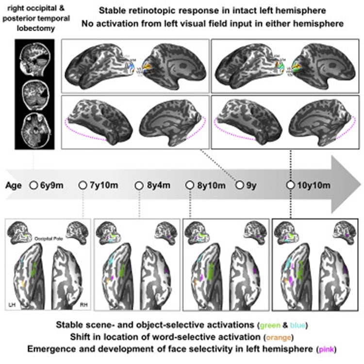
Of the four lobes of UD’s right hemisphere, the entire occipital lobe, which includes the vision processing center of the brain, and most of the temporal lobe, which receives both visual and auditory cues, were removed. His entire left hemisphere was left intact. Because the right half of the brain receives visual information about views to the left, to UD, the left side of the world simply isn’t there.
Despite that, UD performs normally on tasks such as pattern perception. Researchers found that his brain reorganized to compensate for some higher-order functions such as analysis of complex visual cues needed for face and object recognition, but not lower-order functions such as receiving and transmitting all visual aspects of an 180-degree scene.
Behrmann says that, even though roughly a third of UD’s cerebral hemisphere has been removed, his intellect, visual perception, and face and object recognition skills are age appropriate.
“The only deficit is that he could not see the entire visual field–when he is looking forward, visual information falling on the left side of the input is not processed–but he could compensate for this by turning his head or moving his eyes,” Behrmann says. The researchers were surprised that the intact regions of the left hemisphere of UD’s brain came to do “all the work of processing patterns such as faces, objects, and words” for both hemispheres.
“Moreover, by tracking the changes that occurred in the brain as UD developed, we were able to show which parts of the brain remained stable and which were reorganized over time,” she says, offering insight into how the brain can remap visual function in the cortex.
“While we know the human brain has incredible potential to reorganize and remap, it seems that humans still require two intact and functional hemispheres to have a full 180 degrees of peripheral vision without having to move our eyes or turn our heads to avoid blind spots,” Behrmann says. “Our results indicate dynamic reorganization over time in higher-order functions, but not the lower-order visual cortex.”
Lobectomy–the procedure UD underwent–is rare, done on only 4-6 percent of patients of all ages with medically intractable epilepsy. Yet, the surgery completely eliminates seizures for 60-70 percent of children who have it done.
Children who have lost large sections of their brains to disease or injury offer researchers a rare opportunity to elucidate the nature and extent of plasticity–the ability of sections of the brain to take over the the functions of missing or damaged regions.
UD, now almost eleven, is free of seizures. As before the surgery, his IQ is above average and his language skills age appropriate. His test scores are proficient to advanced. At school, he receives vision therapy and sits on the left side of classrooms to allow him to take in more of the scene.
Using functional magnetic resonance imaging (fMRI) to noninvasively measure UD’s brain activity at five different points over three years, the researchers saw regions in charge of face and word recognition emerge later and compete for cortical representation, providing a rare view of plasticity in action.
Behrmann says many provocative scientific questions remain, such as: Which lobectomy patients will show recovery, which will not, and why not? Are patients more likely to regain functions if the left or the right hemisphere is removed? And, is recovery of the visual system more robust in younger individuals?
Funding: This research is supported by the National Institutes of Health.
Source: Carly Britton – Cell Press
Publisher: Organized by NeuroscienceNews.com.
Image Source: NeuroscienceNews.com image is credited to Behrmann et al./Cell Reports.
Original Research: Open access research for “Successful Reorganization of Category-Selective Visual Cortex following Occipito-temporal Lobectomy in Childhood” by Tina T. Liu, Adrian Nestor, Mark D. Vida, John A. Pyles, Christina Patterson, Ying Yang, Fan Nils Yang, Erez Freud, and Marlene Behrmann in Cell Reports. Published May 7 2018.
doi:10.1016/j.celrep.2018.06.099
[cbtabs][cbtab title=”MLA”]Cell Press”Long Term Study of a Boy’s Lobectomy Offers Rare Glimpse of Plasticity in Action.” NeuroscienceNews. NeuroscienceNews, 31 July 2018.
<https://neurosciencenews.com/lobectomy-plasticity-9639/>.[/cbtab][cbtab title=”APA”]Cell Press(2018, July 31). Long Term Study of a Boy’s Lobectomy Offers Rare Glimpse of Plasticity in Action. NeuroscienceNews. Retrieved July 31, 2018 from https://neurosciencenews.com/lobectomy-plasticity-9639/[/cbtab][cbtab title=”Chicago”]Cell Press”Long Term Study of a Boy’s Lobectomy Offers Rare Glimpse of Plasticity in Action.” https://neurosciencenews.com/lobectomy-plasticity-9639/ (accessed July 31, 2018).[/cbtab][/cbtabs]
Abstract
Successful Reorganization of Category-Selective Visual Cortex following Occipito-temporal Lobectomy in Childhood
Investigations of functional (re)organization in children who have undergone large cortical resections offer a unique opportunity to elucidate the nature and extent of cortical plasticity. We report findings from a 3-year investigation of a child, U.D., who underwent surgical removal of the right occipital and posterior temporal lobes at age 6 years 9 months. Relative to controls, post-surgically, U.D. showed age-appropriate intellectual performance and visuoperceptual face and object recognition skills. Using fMRI at five different time points, we observed a persistent hemianopia and no visual field remapping. In category-selective visual cortices, however, object- and scene-selective regions in the intact left hemisphere were stable early on, but regions subserving face and word recognition emerged later and evinced competition for cortical representation. These findings reveal alterations in the selectivity and topography of category-selective regions when confined to a single hemisphere and provide insights into dynamic functional changes in extrastriate cortical architecture.


