Summary: Researchers report the accumulation of glycosphingolipids in the substantia nigra appears to be elevated in Parkinson’s disease patients. The finding could be used as a biomarker for helping diagnose Parkinson’s before symptoms of the disease set in.
Source: McLean Hospital.
A collaborative team of researchers at McLean Hospital, a Harvard Medical School affiliate, and Oxford University has found that elevated levels of certain types of lipids (fat molecules) in the brain may be an early sign of Parkinson’s disease (PD). This finding could have significant implications for identifying patients who may be at risk for developing PD and for the early treatment of the disease. The detailed findings are available in the April 29 online edition of Neurobiology of Aging.
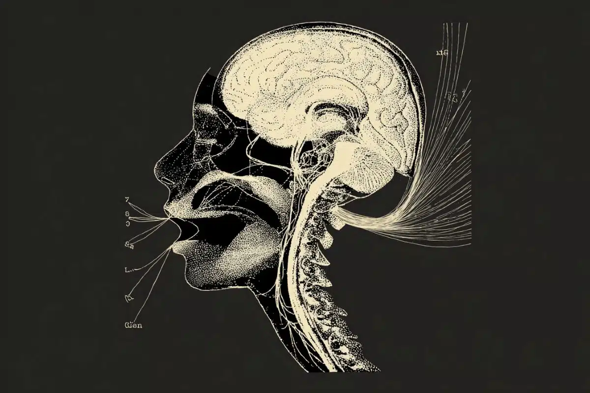
Parkinson’s disease is a degenerative, progressive disorder characterized by the dramatic reduction of nerve cells, particularly dopamine neurons that are involved in movement initiation, in an area of the brain called the substantia nigra. For many years now, the loss of these nerve cells has been attributed to the toxic accumulation of the protein alpha-synuclein. In the past 15 years, however, researchers have been studying an interesting relationship between the risk of developing PD and a group of disorders called lysosomal storage diseases–particularly Gaucher disease, which is caused by mutations that lead to loss of function in the glucocerebrosidase (GBA) gene.
The GBA gene normally produces an enzyme that breaks down lipids, but in the childhood disorder Gaucher disease, a near total lack of this enzyme activity leads to massive and usually fatal elevations of lipids inside cells. Notably, people who do not develop Gaucher disease, but are carriers of one defective gene copy, have a 7 to 10-fold risk of developing PD with age.
“This means that lipid accumulation may also be important in PD, and scientists at the Neuroregeneration Research Institute at McLean Hospital have previously shown that there is an elevation of a class of lipids, called glycosphingolipids, in the substantia nigra of patients with PD,” said Ole Isacson, MD, PhD, professor of Neurology and Neuroscience at Harvard Medical School, co-director of the Neuroregeneration Research Institute at McLean Hospital, and co-senior author of the study.
Since aging is the most significant risk factor for developing PD, teams from McLean Hospital and the University of Oxford laboratory of Professor Frances M. Platt, PhD, FMedSci, collaborated to measure the levels of glycosphingolipids in the aging brain, using young and old mice. They found that the same glycosphingolipids that are increased in the brains of Parkinson’s disease patients are also elevated in the brains of aging mice. These findings show that both genetics (GBA gene mutation) and aging can cause the same lipid elevations in the brain that are demonstrated in Parkinson’s disease pathology.
“These results lead to a new hypothesis that lipid alterations may create a number of problems inside nerve cells in degenerative aging and Parkinson’s disease, and that these changes may precede some of the more obvious hallmarks of Parkinson’s disease, such as protein aggregates,” said Penny Hallett, PhD, study lead author and co-director of McLean’s Neuroregeneration Research Institute. “This potentially provides an opportunity to treat lipid changes early on in Parkinson’s disease and protect nerve cells from dying, as well as the chance to use the lipid levels as biomarkers for patients at risk.”
Funding: This work was funded by the National Institutes of Health, Neuroregeneration Institute at McLean Hospital, Parkinson’s UK, Wellcome Trust.
Source: Laura Neves – McLean Hospital
Publisher: Organized by NeuroscienceNews.com.
Image Source: NeuroscienceNews.com image is credited to Stockstill et al., 2018.
Original Research: Open access research for “Glycosphingolipid levels and glucocerebrosidase activity are altered in normal aging of the mouse brain” by Penelope J. Hallett, Mylene Huebecker, Oeystein R. Brekk, Elizabeth B. Moloney, Emily M. Rocha, David A. Priestman, Frances M. Platt, and Ole Isacson in Neurobiology of Aging. Published March 29 2018.
doi:10.1016/j.neurobiolaging.2018.02.028
[cbtabs][cbtab title=”MLA”]McLean Hospital “Lipid Accumulation in Brain May Be an Early Sign of Parkinson’s.” NeuroscienceNews. NeuroscienceNews, 29 April 2018.
<https://neurosciencenews.com/lipid accumulation-parkinsons-8912/>.[/cbtab][cbtab title=”APA”]McLean Hospital (2018, April 29). Lipid Accumulation in Brain May Be an Early Sign of Parkinson’s. NeuroscienceNews. Retrieved April 29, 2018 from https://neurosciencenews.com/lipid accumulation-parkinsons-8912/[/cbtab][cbtab title=”Chicago”]McLean Hospital “Lipid Accumulation in Brain May Be an Early Sign of Parkinson’s.” https://neurosciencenews.com/lipid accumulation-parkinsons-8912/ (accessed April 29, 2018).[/cbtab][/cbtabs]
Abstract
Glycosphingolipid levels and glucocerebrosidase activity are altered in normal aging of the mouse brain
Aging is the predominant risk factor for both genetic and sporadic Parkinson’s disease (PD). The majority of PD cases are nonfamilial, and the connection between aging and PD-associated genes is not well understood. Haploinsufficiency of the GBA gene, leading to a reduction in glucocerebrosidase (GCase) activity, is one of the most common genetic risk factors for PD. Furthermore, GCase activity is also reduced in brain regions of sporadic PD patients, with a corresponding accumulation of its glycosphingolipid (GSL) substrates. Recent findings in PD patients and aging control cases, and in human PD patient induced pluripotent stem cell neurons, have shown an age-dependent reduction in GCase activity and an elevation of some GSLs. We therefore asked whether aging-induced changes to both lysosomal and nonlysosomal GCase activity and GSL homeostasis in the brain could also be reflected in other nonhuman mammalian systems. Increases in brain polyubiquitin and the lysosomal-associated membrane protein, LAMP2A, were found in 24-month-old wild-type mice compared to 1.5-month-old mice. A lipidomic analysis was performed on brains of wild-type mice of different strains between 1.5 and 24 months of age. Aging created GSL changes that are reminiscent of sporadic PD. Levels of glucosylceramide, glucosylsphingosine, lactosylceramide, and GM1a were elevated in the brain of aged mice, and levels of complex gangliosides, GD1a, GD1b, and GT1b, were reduced with age. Parallel biochemical analyses revealed a change in lipid metabolism probably mediated by lysosomal hydrolases, with reduced GCase and increased neuraminidase activity. Based on these data, we hypothesize that perturbation of GSL metabolism in the aging brain may precede or may be part of abnormal protein handling and may accelerate PD pathophysiological processes in vulnerable neurons in PD and other age-related neurodegenerative disorders.