

Summary:
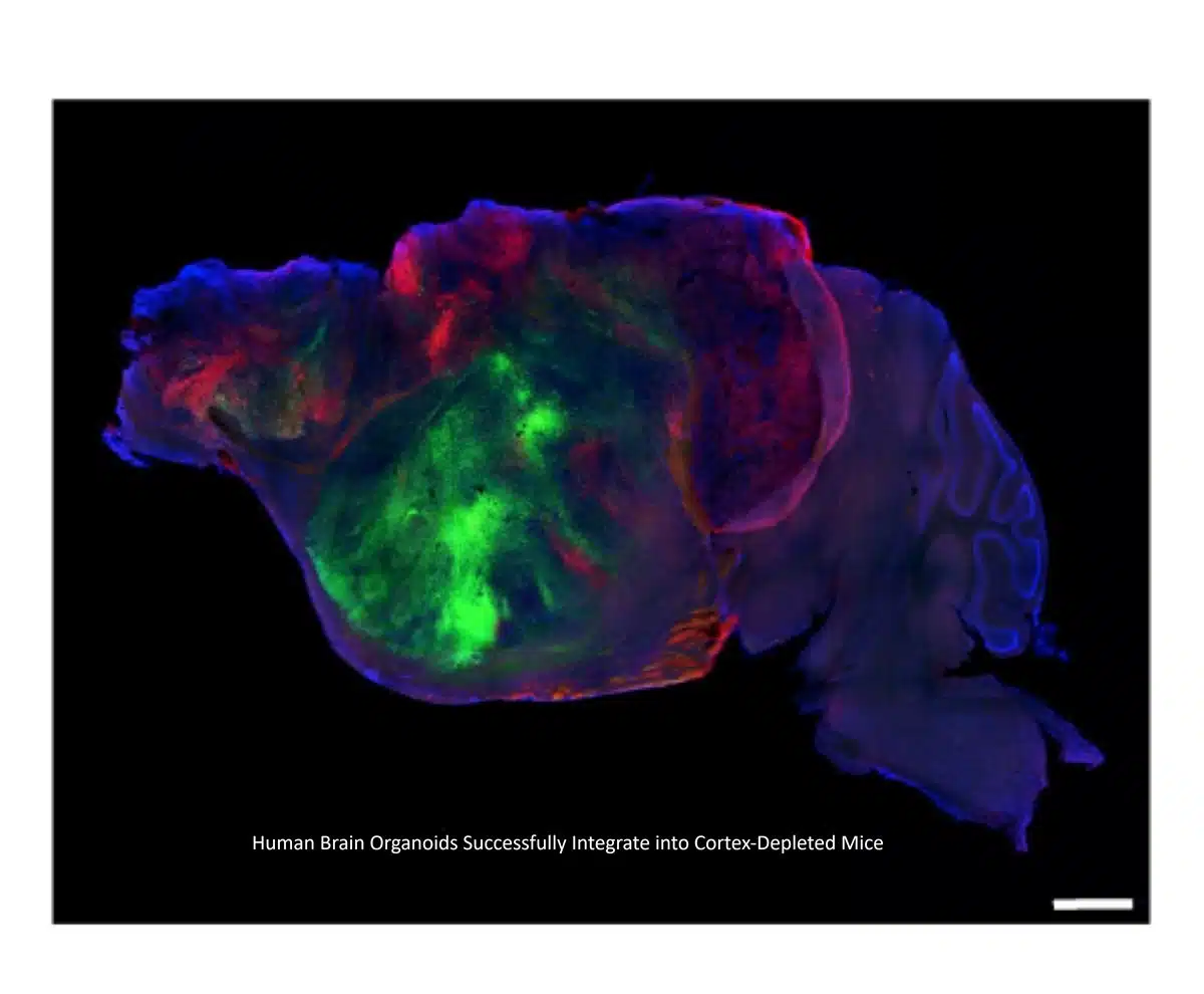
Neuroscientists have developed a genetic strategy to produce mice lacking most of their cerebral cortex, creating the physical space needed for transplanted human brain organoids to expand, integrate, and mature. The engrafted human tissue differentiated into diverse neuronal and glial populations, including cell types previously unattainable in vitro, and formed functional, circuit-level connections with the host brain and spinal cord that influenced behavior.
Key Facts:
- Cortex Depletion Creates Structural Room: To overcome cranial space constraints and competition with host neurons, researchers genetically engineered mice to develop without the vast majority of their cerebral cortex, allowing human organoids to occupy and rebuild cortical structures.
- Cellular Diversity and In Vivo Maturation: The grafted human cells differentiated into an array of specialized brain cells, including rare cell types that fail to mature in conventional laboratory dish cultures, and established functional neural networks reaching down to the mouse spinal cord.
- Behavioral and Disease Modeling Impact: Organoid-engrafted mice exhibited measurable differences in motor and memory tasks compared to non-transplanted controls, providing a physiological platform to model disorders like hypoxic brain injury and neurodevelopmental conditions.
Source: Stanford University / Nature
Investigating the nuanced mechanisms of human brain development, psychiatric conditions, and neurodegenerative disorders has long been constrained by the physical inaccessibility of living human cortical tissue. While 3D stem-cell-derived brain organoids offer an unprecedented window into cellular pathology, growing them in laboratory culture dishes limits their lifespan, vascularization, and circuit maturation.
Prior attempts to transplant human organoids into rodent models offered a partial solution, but the tissue invariably collided with physical bottlenecks: rigid cranial borders and competitive suppression by dense rodent neural architecture prevented the human grafts from achieving full morphological and functional scale.
Now, in a study published in Nature, a research team led by Dr. Sergiu Pașca has bypassed this barrier through developmental engineering. By genetically engineering mice to develop with severely reduced cortical volume, essentially missing the outer rind of the cerebral hemispheres, the investigators carved out a physiological niche for human brain organoids to settle, vascularize, and construct intricate, working neural systems.
Cellular Differentiation Beyond the Petri Dish
Following transplantation into the cortex-depleted cavity, the human brain organoids flourished. The tissue expanded to fill the void, organizing into stratified layers and complex microarchitectures that closely mirrored authentic human cortical tissue.
Critically, the in vivo environment provided biological cues that standard incubators cannot replicate. The grafts generated a wide variety of human neurons and glia, including distinct neural subtypes that scientists have historically struggled to culture in vitro.
Rather than existing as isolated cellular colonies, the human neurons extended projection axons throughout the host central nervous system. Electrophysiological and anatomical tracings confirmed that the human grafts wired functional synaptic connections into the host mouse brainstem and descended into the spinal cord, bridging human cellular machinery with rodent motor execution networks.
Behavioral Rescue and Disease Modeling
To test whether the engrafted human cortex influenced functional outputs, the researchers evaluated the animals across a battery of behavioral paradigms. Mice carrying the human organoid grafts displayed marked differences in motor coordination and memory performance compared to cortex-depleted mice that did not receive transplants.
The team also demonstrated the platform’s utility as an experimental disease testbed. By exposing the chimeric models to hypoxic injury, a deprivation of oxygen that underlies perinatal stroke and cerebral palsy, the researchers observed subtle, measurable changes in gait and locomotion. This confirmed that the hybrid platform can translate cellular-level insults within human tissue into clear, behavioral readouts in a living organism.
Navigating the Ethics of In Vivo Brain Models
Because the platform blurs traditional boundaries between human cellular architecture and animal physiology, the researchers underscored that all experiments adhered strictly to rigorous, specialized ethical protocols.
The authors called for proactive, ongoing discussions among scientists, bioethicists, and regulatory bodies to formalize guidelines addressing the societal, clinical, and ethical nuances of brain organoid chimeras. Looking forward, the team hopes the system will accelerate therapies for intractable neurodevelopmental disorders by testing prospective drugs directly against maturing human neural circuits in a living host.
Editorial Notes:
- This article was edited by a Neuroscience News editor.
- Journal paper reviewed in full.
- Additional context added by our staff.
About this Genetics and Neurology Research:
- Media Contact: Sergiu Pașcal
- Source: Nature
- Image Credit: Image credited to S. Pasca lab, Stanford University/Nature
- Original Research is Open Access: Nature (September 16, 2026). “Developmental xenocortication using human-derived organoids in mice” Authors: Konstantin Kaganovsky, Kevin W. Kelley, Tilo Gschwind, Paul M. Harary, John Kochalka, Alexander D. White, Garikoitz Lerma-Usabiaga, Xiaoyu Chen, Omer Revah, Felicity Gore, Ayano Aoyama, Jennifer L. Shadrach, Se-Jin Yoon, Alfredo Valencia, Satoe Ogawa, Noah Reis, Hannes Vogel, Brian Wandell, Julia A. Kaltschmidt, Ivan Soltesz, Karl Deisseroth & Sergiu P. Pașca.
- DOI: 10.1038/s41586-026-11032-2
Abstract
Developmental xenocortication using human-derived organoids in mice
The inaccessibility of human brain tissue limits the study of human development and function, a challenge that human stem-cell-derived neural models are beginning to address. Transplantation of neural organoids into rodent hosts enables the in vivo study of aspects of human neurodevelopment and circuit function, alongside behavioural phenotyping of the host animals.
However, spatial limitations and competition with host circuits constrain the integration of neural organoids, which is critical for studying disease.
Here we establish a transplantation platform using a genetic strategy to effectively deplete glutamatergic neurons from mouse neocortex and hippocampus (apallial) and neonatally engraft the cortical cavity with human stem-cell-derived cortical organoids (hCO) to generate xenocortical mice. This leads to robust graft growth with hCOs occupying most of the cortical volume and generating a diversity of human cortical cell types, including layer 5 extratelencephalic projection neurons.
Human cortical neurons integrate with the mouse nervous system, and in vivo cortical graft-wide calcium imaging and electrophysiological analyses revealed patterns of organized activity resembling developing circuits. Behavioural analyses of apallial and xenocortical mice revealed broadly preserved locomotion alongside selective differences in limb coordination and altered organization of spontaneous behaviour.
Lastly, this platform enabled behavioural readouts in a model of injury to developing human cortical cells. We envision that xenocortication will be useful for obtaining circuit- and behaviour-level readouts using human neurons to study neurodevelopment, model disease and develop therapeutics.


