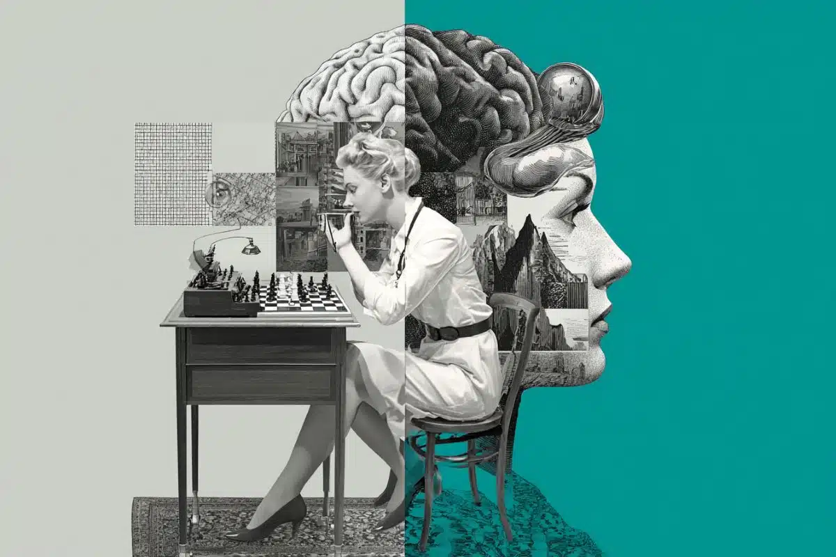
UC San Francisco scientists have discovered a network of brain cells that allows animals to keep track of where they are when they are not moving through space, such as when they are eating, engaged in social interactions, or sleeping. The new research fills a critical gap in a decades-long line of research on the so-called GPS system of the brain, which has so far been largely described in terms of cells that aid navigation by becoming active when animals, including humans, move through particular locations.
“Experiences when you’re moving are different from experiences when you’re still,” said Loren Frank, PhD, professor of physiology at UCSF and senior author of the new study, which appears in the March 2 online issue of Nature. “When you’re creating memories of a trip you’re taking, there are memories of all the different places you’ve passed through, but there is a whole other class of experiences you have when you’re just in one place, and our work shows that the brain actually processes these experiences in a different way.”
Frank is a Howard Hughes Medical Institute (HHMI) investigator, and co-director of the Kavli Institute for Fundamental Neuroscience at UCSF.
In 2014, the Nobel Prize in Physiology or Medicine recognized the discoveries of the brain’s “place cells” and “grid cells,” which together comprise a cognitive mapping system that animals rely on to navigate. Place cells, which are found in the brain’s hippocampus, a structure crucial to memory, fire robustly when animals pass through particular spatial locations. Grid cells, in the entorhinal cortex, fire like place cells but in a repeating pattern of spatial locations.
In recent work, Frank and others have shown that, when animals aren’t moving, they occasionally “replay” earlier sequences of place-cell firing, perhaps as mental rehearsal for long-term storage or as a way to plan out future locations to visit. But because place cells were thought to be active mainly when animals walk or run, it was not clear whether or how animals kept track of location when not moving.
To explore this question, a team led by UCSF graduate student Kenneth Kay used a W-shaped maze containing a reward at three different sites, then trained rats to travel among the three sites in a particular order. This study design ensured that the animals would experience not only periods of movement when in the maze, but also periods of stillness when receiving rewards and when considering where to go next in the maze.
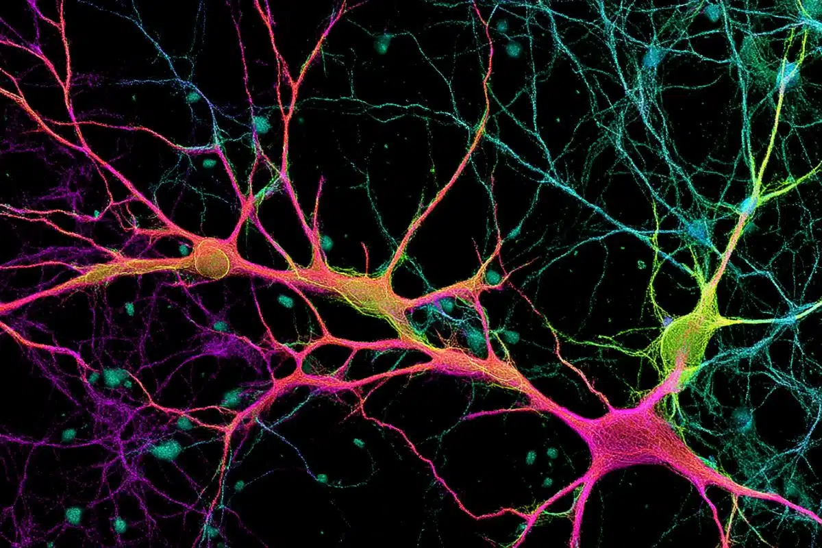
The scientists recorded activity in four different hippocampal regions and found that cells in an area called CA2 became especially active when the rats ceased moving, and that these cells were part of a wider network of hippocampal cells that signal rats’ location during immobile periods. Signaling of location was even found to be active while the rats were sleeping.
Interestingly, the signal for current location was found to turn off rapidly during place-cell replay and vice versa, Frank said, suggesting that the hippocampus switches between representing past experience versus current experience, on a timescale of tenths of a second.
“Some research has suggested that CA2 is important in forming social memories, which is intriguing, because social experience in rats generally takes place when they’re not moving around a lot,” Frank said. “The same can be said for our own social experiences. We can go biking with someone, but a lot of these experiences take place when we’re just sitting together. It may be that a system that’s specialized for knowing where you are and what the context is when you’re not moving could be important for remembering social interactions.”
Funding: The research was supported by HHMI, the National Institutes of Health, and a McKnight Foundation award to Frank, and a Ruth L. Kirchstein National Research Service Award Fellowship to Kay, a student in the UCSF Medical Scientist Training Program.
Source: Pete Farley – UCSF
Image Credit: The image is adapted from the UCSF press release.
Original Research: Abstract for “A hippocampal network for spatial coding during immobility and sleep” by Kenneth Kay, Marielena Sosa, Jason E. Chung, Mattias P. Karlsson, Margaret C. Larkin and Loren M. Frank in Nature. Published online March 2 2016 doi:10.1038/nature17144
Abstract
A hippocampal network for spatial coding during immobility and sleep
How does an animal know where it is when it stops moving? Hippocampal place cells fire at discrete locations as subjects traverse space, thereby providing an explicit neural code for current location during locomotion. In contrast, during awake immobility, the hippocampus is thought to be dominated by neural firing representing past and possible future experience. The question of whether and how the hippocampus constructs a representation of current location in the absence of locomotion has been unresolved. Here we report that a distinct population of hippocampal neurons, located in the CA2 subregion, signals current location during immobility, and does so in association with a previously unidentified hippocampus-wide network pattern. In addition, signalling of location persists into brief periods of desynchronization prevalent in slow-wave sleep. The hippocampus thus generates a distinct representation of current location during immobility, pointing to mnemonic processing specific to experience occurring in the absence of locomotion.
“A hippocampal network for spatial coding during immobility and sleep” by Kenneth Kay, Marielena Sosa, Jason E. Chung, Mattias P. Karlsson, Margaret C. Larkin and Loren M. Frank in Nature. Published online March 2 2016 doi:10.1038/nature17144