Researchers point to dietary link for proper pre- and postnatal neural growth.
While recent reports question whether fish oil supplements support heart health, UC Irvine scientists have found that the fatty acids they contain are vitally important to the developing brain.
In a study appearing today in the Journal of Neuroscience, UCI neurobiologists report that dietary deficiencies in the type of fatty acids found in fish and other foods can limit brain growth during fetal development and early in life. The findings suggest that women maintain a balanced diet rich in these fatty acids for themselves during pregnancy and for their babies after birth.
Susana Cohen-Cory, professor of neurobiology & behavior, and colleagues identified for the first time how deficits in what are known as n-3 polyunsaturated fatty acids cause molecular changes in the developing brain that result in constrained growth of neurons and the synapses that connect them.
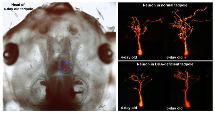
These fatty acids are precursors of docosahexaenoic acid, or DHA, which plays a key role in the healthy creation of the central nervous system. In their study, which used female frogs and tadpoles, the UCI researchers were able to see how DHA-deficient brain tissue fostered poorly developed neurons and limited numbers of synapses, the vital conduits that allow neurons to communicate with each other.
“Additionally, when we changed the diets of DHA-deficient mothers to include a proper level of this dietary fatty acid, neuronal and synaptic growth flourished and returned to normal in the following generation of tadpoles,” Cohen-Cory said.
DHA is essential for the development of a fetus’s eyes and brain, especially during the last three months of pregnancy. It makes up 10 to 15 percent of the total lipid amount of the cerebral cortex. DHA is also concentrated in the light-sensitive cells at the back of the eyes, where it accounts for as much as 50 percent of the total lipid amount of each retina.
Dietary DHA is mainly found in animal products: fish, eggs and meat. Oily fish – mackerel, herring, salmon, trout and sardines – are the richest dietary source, containing 10 to 100 times more DHA than nonmarine foods such as nuts, seeds, whole grains and dark green, leafy vegetables.
DHA is also found naturally in breast milk. Possibly because of this, the fatty acid is used as a supplement for premature babies and as an ingredient in baby formula during the first four months of life to promote better mental development.
The UCI team utilized Xenopus laevis (the African clawed frog) as a model for this study because it allowed them to follow the progression and impact of the maternal dietary deficit in the offspring. Because frog embryos develop outside the mother and are translucent, the researchers could see dynamic changes in neurons and their synaptic connections in the intact, live embryos, where development can be easily studied from the time of fertilization to well after functional neural circuits form.
They focused on the visual system because it’s an accessible and well-established system known to depend on fatty acids for proper growth and utility.
Miki Igarashi and Rommel Santos of UC Irvine contributed to the study.
Funding: The research was funded by the National Eye Institute (grant EY-011912).
Source: UC Irvine
Image Source: The image is credited to Cohen-Cory lab / UC Irvine
Original Research: Abstract for “Impact of Maternal n-3 Polyunsaturated Fatty Acid Deficiency on Dendritic Arbor Morphology and Connectivity of Developing Xenopus laevis Central Neurons In Vivo” by Miki Igarashi, Rommel A. Santos, and Susana Cohen-Cory in Journal of Neuroscience. Published online April 15 2015 doi:10.1523/JNEUROSCI.4102-14.2015
Abstract
Impact of Maternal n-3 Polyunsaturated Fatty Acid Deficiency on Dendritic Arbor Morphology and Connectivity of Developing Xenopus laevis Central Neurons In Vivo
Docosahexaenoic acid (DHA, 22:6n-3) is an essential component of the nervous system, and maternal n-3 polyunsaturated fatty acids (PUFAs) are an important source for brain development. Here, the impact of DHA on developing central neurons was examined using an accessible in vivo model. Xenopus laevis embryos from adult female frogs fed n-3 PUFA-adequate or deficient diets were analyzed every 10 weeks for up to 60 weeks, when frogs were then switched to a fish oil-supplemented diet. Lipid analysis showed that DHA was significantly reduced both in oocytes and tadpoles 40 weeks after deprivation, and brain DHA was reduced by 57% at 60 weeks. In vivo imaging of single optic tectal neurons coexpressing tdTomato and PSD-95-GFP revealed that neurons were morphologically simpler in tadpoles from frogs fed the deficient diet compared with the adequate diet. Tectal neurons had significantly fewer dendrite branches and shorter dendritic arbor over a 48 h imaging period. Postsynaptic cluster number and density were lower in neurons deprived of n-3 PUFA. Moreover, changes in neuronal morphology correlated with a 40% decrease in the levels of BDNF mRNA and mature protein in the brain, but not in TrkB. Importantly, switching to a fish oil-supplemented diet induced a recovery in DHA content in the frog embryos within 20 weeks and diminished the deprivation effects observed on tectal neurons of Stage 45 tadpoles. Consequently, our results indicate that DHA impacts dendrite maturation and synaptic connectivity in the developing brain, and it may be involved in neurotrophic support by BDNF.
“Impact of Maternal n-3 Polyunsaturated Fatty Acid Deficiency on Dendritic Arbor Morphology and Connectivity of Developing Xenopus laevis Central Neurons In Vivo” by Miki Igarashi, Rommel A. Santos, and Susana Cohen-Cory in Journal of Neuroscience. Published online April 15 2015 doi:10.1523/JNEUROSCI.4102-14.2015