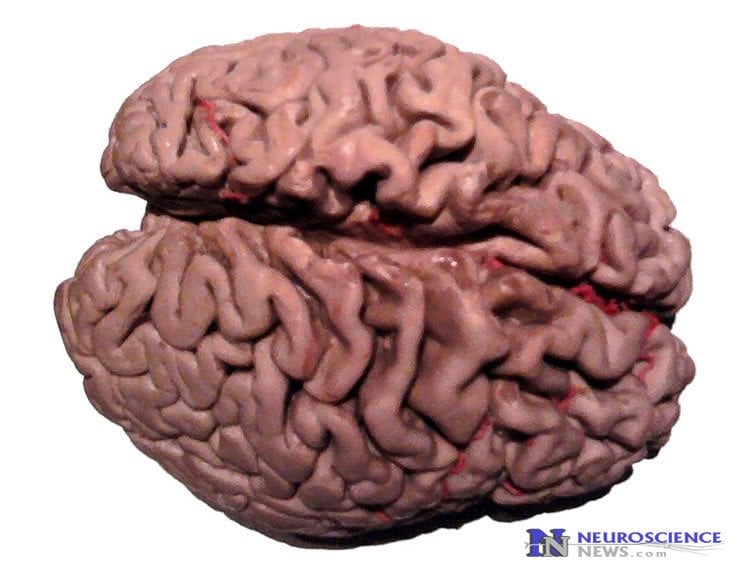
New research has revealed how disease-associated changes in 2 interlinked networks within the brain may play a key role in the development of the symptoms of dementia.
The University of Exeter Medical School led two studies, each of which moves us a step closer to understanding the onset of dementia, and potentially to paving the way for future therapies. Both studies, part-funded by Alzheimer’s Research UK, are published in the Journal of Neuroscience and involved collaboration with the University of Bristol.
Both studies shed light on how two parts of the brain’s ‘GPS’ navigation system malfunctions in dementia, and point to likely underpinning causes for loss of orientation that is commonly experienced by people living with the condition.
In the first study, the team studied a part of the brain called the entorhinal cortex. Located near the base of the brain, this region is associated with functions including memory formation and navigation, and contains so-called “grid cells”. These nerve cells fire electrical discharges in a grid-like pattern, much like the grid on an Ordnance Survey map. Paralleling the different scales employed by different maps, the grid firing patterns in the entorhinal cortex also have different scales, with cells at the top of the cortex having a more tightly packed grid pattern than those at the bottom. Scientists believe that this top-to-bottom gradient of different grid scales contributes pivotally to our sense of spatial location.
The team compared the activity in the entorhinal cortex of healthy mice and mice with dementia. They found that top-to-bottom gradients in electrical activity in the entorhinal cortex are not present in mice with dementia. Their findings suggest that the fine navigational detail, such as you would find on a large-scale map, is not correctly represented in patients with dementia.
Dr. Jon Brown at the University of Exeter Medical School led the studies, as part of his Alzheimer’s Research UK Senior Fellowship. He said: “This is an exciting discovery because it is the first time grid cell activity has been linked to the onset of disease. We now need further research to better establish how these findings translate to dementia in humans.”
In the second study, researchers examined “place cells” located in the hippocampus, a brain structure known to be critical in processing learning and memory, both affected by dementia. Place cells help us to identify where we are within a certain space.
The team found that the hippocampus of mice with dementia was associated with specific disturbances in synaptic, cellular, and network-level function, meaning that spatial information was wrongly encoded and spatial memory was impaired.

Dr. Brown said: “Dementia is one of the greatest health challenges of our time, and we still have so much to learn about its causes, as well as about how our brains work. This research makes progress in both areas, and is another small step along the road to earlier diagnoses and finding new treatments and therapies.”
Professor Andrew Randall, who co-supervised much of the work, said: “This has been a fascinating experimental journey for our research teams, and much of the pivotal work was carried out by talented PhD students. We look forward to producing much more work of this nature as members of Exeter’s growing dementia research community.”
Dr. Laura Phipps from Alzheimer’s Research UK, said: “There are 850,000 people in the UK with dementia and a tenth of those are living in the South West. It is vital that researchers explore the complexities of the brain, to understand more about the causes of the condition and how we can tackle it. Dementia is not just a synonym for forgetfulness – these findings in mice highlight the impact that diseases like Alzheimer’s can have on spatial orientation. It will now be important to build on this research, to understand whether this chain of events can be targeted in the hunt for new treatments.”
The University of Exeter forms part of the Alzheimer’s Research UK South West Research Network – a community of dementia researchers in Exeter and Plymouth, working collaboratively to accelerate progress in dementia research.
Funding: Funding was provided by Alzheimer’s Research UK.
Source: Louise Vennells – University of Exeter
Image Source: The image is in the public domain
Original Research: Full open access research for for “Electrical and Network Neuronal Properties Are Preferentially Disrupted in Dorsal, But Not Ventral, Medial Entorhinal Cortex in a Mouse Model of Tauopathy” by Clair A. Booth, Thomas Ridler, Tracey K. Murray, Mark A. Ward, Emily de Groot, Marc Goodfellow, Keith G. Phillips, Andrew D. Randall, and Jonathan T. Brown in Journal of Neuroscience. Published online January 13 2016 doi:10.1523/JNEUROSCI.2845-14.2016
Full open access research for for “Altered Intrinsic Pyramidal Neuron Properties and Pathway-Specific Synaptic Dysfunction Underlie Aberrant Hippocampal Network Function in a Mouse Model of Tauopathy” by Clair A. Booth, Jonathan Witton, Jakub Nowacki, Krasimira Tsaneva-Atanasova, Matthew W. Jones, Andrew D. Randall, and Jonathan T. Brown in Journal of Neuroscience. Published online January 13 2016 doi:10.1523/JNEUROSCI.2151-15.2016
Abstract
Electrical and Network Neuronal Properties Are Preferentially Disrupted in Dorsal, But Not Ventral, Medial Entorhinal Cortex in a Mouse Model of Tauopathy
The entorhinal cortex (EC) is one of the first areas to be disrupted in neurodegenerative diseases such as Alzheimer’s disease and frontotemporal dementia. The responsiveness of individual neurons to electrical and environmental stimuli varies along the dorsal–ventral axis of the medial EC (mEC) in a manner that suggests this topographical organization plays a key role in neural encoding of geometric space. We examined the cellular properties of layer II mEC stellate neurons (mEC-SCs) in rTg4510 mice, a rodent model of neurodegeneration. Dorsoventral gradients in certain intrinsic membrane properties, such as membrane capacitance and afterhyperpolarizations, were flattened in rTg4510 mEC-SCs, while other cellular gradients [e.g., input resistance (Ri), action potential properties] remained intact. Specifically, the intrinsic properties of rTg4510 mEC-SCs in dorsal aspects of the mEC were preferentially affected, such that action potential firing patterns in dorsal mEC-SCs were altered, while those in ventral mEC-SCs were unaffected. We also found that neuronal oscillations in the gamma frequency band (30–80 Hz) were preferentially disrupted in the dorsal mEC of rTg4510 slices, while those in ventral regions were comparatively preserved. These alterations corresponded to a flattened dorsoventral gradient in theta-gamma cross-frequency coupling of local field potentials recorded from the mEC of freely moving rTg4510 mice. These differences were not paralleled by changes to the dorsoventral gradient in parvalbumin staining or neurodegeneration. We propose that the selective disruption to dorsal mECs, and the resultant flattening of certain dorsoventral gradients, may contribute to disturbances in spatial information processing observed in this model of dementia.
SIGNIFICANCE STATEMENT The medial entorhinal cortex (mEC) plays a key role in spatial memory and is one of the first areas to express the pathological features of dementia. Neurons of the mEC are anatomically arranged to express functional dorsoventral gradients in a variety of neuronal properties, including grid cell firing field spacing, which is thought to encode geometric scale. We have investigated the effects of tau pathology on functional dorsoventral gradients in the mEC. Using electrophysiological approaches, we have shown that, in a transgenic mouse model of dementia, the functional properties of the dorsal mEC are preferentially disrupted, resulting in a flattening of some dorsoventral gradients. Our data suggest that neural signals arising in the mEC will have a reduced spatial content in dementia.
“Electrical and Network Neuronal Properties Are Preferentially Disrupted in Dorsal, But Not Ventral, Medial Entorhinal Cortex in a Mouse Model of Tauopathy” by Clair A. Booth, Thomas Ridler, Tracey K. Murray, Mark A. Ward, Emily de Groot, Marc Goodfellow, Keith G. Phillips, Andrew D. Randall, and Jonathan T. Brown in Journal of Neuroscience. Published online January 13 2016 doi:10.1523/JNEUROSCI.2845-14.2016
Abstract
Altered Intrinsic Pyramidal Neuron Properties and Pathway-Specific Synaptic Dysfunction Underlie Aberrant Hippocampal Network Function in a Mouse Model of Tauopathy
The formation and deposition of tau protein aggregates is proposed to contribute to cognitive impairments in dementia by disrupting neuronal function in brain regions, including the hippocampus. We used a battery of in vivo and in vitro electrophysiological recordings in the rTg4510 transgenic mouse model, which overexpresses a mutant form of human tau protein, to investigate the effects of tau pathology on hippocampal neuronal function in area CA1 of 7- to 8-month-old mice, an age point at which rTg4510 animals exhibit advanced tau pathology and progressive neurodegeneration. In vitro recordings revealed shifted theta-frequency resonance properties of CA1 pyramidal neurons, deficits in synaptic transmission at Schaffer collateral synapses, and blunted plasticity and imbalanced inhibition at temporoammonic synapses. These changes were associated with aberrant CA1 network oscillations, pyramidal neuron bursting, and spatial information coding in vivo. Our findings relate tauopathy-associated changes in cellular neurophysiology to altered behavior-dependent network function.
SIGNIFICANCE STATEMENT Dementia is characterized by the loss of learning and memory ability. The deposition of tau protein aggregates in the brain is a pathological hallmark of dementia; and the hippocampus, a brain structure known to be critical in processing learning and memory, is one of the first and most heavily affected regions. Our results show that, in area CA1 of hippocampus, a region involved in spatial learning and memory, tau pathology is associated with specific disturbances in synaptic, cellular, and network-level function, culminating in the aberrant encoding of spatial information and spatial memory impairment. These studies identify several novel ways in which hippocampal information processing may be disrupted in dementia, which may provide targets for future therapeutic intervention.
“Altered Intrinsic Pyramidal Neuron Properties and Pathway-Specific Synaptic Dysfunction Underlie Aberrant Hippocampal Network Function in a Mouse Model of Tauopathy” by Clair A. Booth, Jonathan Witton, Jakub Nowacki, Krasimira Tsaneva-Atanasova, Matthew W. Jones, Andrew D. Randall, and Jonathan T. Brown in Journal of Neuroscience. Published online January 13 2016 doi:10.1523/JNEUROSCI.2151-15.2016


