

Summary: Researchers use an experimental PET scan tracer to effectively find brain protein deposits specific to CTE in the brain of a retired NFL player.
Source: Mount Sinai Health System.
Protein tracer shows distinctive pattern of brain protein deposition specific to this disease and typically confirmed after death.
An experimental positron emission tomography (PET) tracer can effectively diagnose concussion-related brain degeneration while a person is still alive, according to a proof-of-concept study conducted at the Icahn School of Medicine at Mount Sinai and published September 27 in the journal Translational Psychiatry.
Mount Sinai researchers used an experimental imaging agent called [18 F]-T807 (or Avid 1451) with PET to examine the brain of a living, 39-year-old retired National Football League (NFL) player who had experienced 22 concussions and exhibited clinical symptoms consistent with chronic traumatic encephalopathy (CTE), a neurodegenerative brain disease that has been associated with repetitive blows to the head in athletes and soldiers. T807 is designed to latch onto a protein called tau that accumulates in the brain as a result of repetitive traumatic brain injury. When the new imaging agent (or ligand) lights up a PET scan of the brain of a patient showing buildup of tau in a characteristic pattern, the scan result is interpreted as being consistent with CTE. Until now, evidence for CTE pathology has only been possible by examining brain tissue after death.
CTE has a distinctive pattern of tau deposition that was described in 2015 by an expert panel commissioned by the National Institute of Neurological Disorders and Stroke (NINDS). That panel laid out diagnostic criteria for CTE based on samples of postmortem brain tissue. The surface of the brain is highly wrinkled. The tau accumulation in CTE appears to trace the highly folded surface of the brain and is especially concentrated at the deepest points in the wrinkles and folds. The NINDS panel used the word “pathognomonic” to describe the CTE tau pathology pattern. This is a technical term that indicates that whenever you see this pattern of tau pathology, the diagnosis can be nothing other than CTE. There can be no confusion with other tau diseases.
“Our study participant’s scan is the first to reveal during life a pattern of tau imaging that outlines the wrinkles and folds of the living brain, just like the ‘pathognomonic pattern’ described by the NINDS panel as diagnostic of a brain with CTE,” says Sam Gandy, MD, Director of the Center for Cognitive Health and NFL Neurological Care Program at the Icahn School of Medicine at Mount Sinai and last author of the study. “When fully validated, this new ligand has the potential to be used as a diagnostic biomarker and represents an exciting development in the detection and tracking of CTE.”
A link between brain injury and long-term health has gained greater attention in recent years, helped along by evidence of neurofibrillary tangles of tau protein, or tauopathy, that has been clinically confirmed in the postmortem brain tissue of former athletes and soldiers with histories of multiple head traumas. In addition to symptoms such as irritability and extreme mood swings, CTE is associated with the symptoms of various other neurodegenerative diseases, including Alzheimer’s, Parkinson’s and Lou Gehrig’s diseases.
“This research is in its infancy,” says Dara L. Dickstein, PhD, Assistant Professor of Neuroscience, and Geriatrics and Palliative Medicine at the Icahn School of Medicine at Mount Sinai and first author of the study. “Whether or not the pathology can be reversed or halted is something we have yet to determine and these new tauopathy PET scans may be able to help in this endeavor.”
Under the leadership of Drs. Gandy and Dickstein, and with primary funding support from the Alzheimer’s Drug Discovery Foundation (ADDF), Mount Sinai is one of the few medical centers researching the use of the new ligand in living patients who are believed to have CTE. The Mount Sinai team is currently studying 24 patients and plans to establish a clinical trial early next year that will employ the new ligand to identify CTE patients who might respond to an anti-tauopathy medicine that is currently being studied at other medical centers for the treatment of Alzheimer’s disease and other neurodegenerative disorders.
“These findings demonstrate that we may now have the first biomarker for the detection of CTE through tau imaging,” says Howard Fillit, MD, ADDF’s Founding Executive Director and Chief Science Officer. “This may prove significant as an early diagnostic tool for those who suffer repeated traumatic brain injuries. It may also help us better understand the similarities in disease processes between CTE, Alzheimer’s and other neurodegenerative diseases, and determine whether repeated head injuries may lead to the onset of Alzheimer’s.”
Funding: In addition to ADDF, this research was supported by the National Institute of Neurological Disorders and Stroke, the National Institute of Child Health and Human Development, Avid Radiopharmaceuticals, Eli Lilly and Company, and the Werber Family Foundation.
Source: Elizabeth Dowling – Mount Sinai Health System
Image Source: This NeuroscienceNews.com image is credited to the researchers/Translational Psychiatry.
Original Research: Full open access research for “Cerebral [18 F]T807/AV1451 retention pattern in clinically probable CTE resembles pathognomonic distribution of CTE tauopathy” by D L Dickstein, M Y Pullman, C Fernandez, J A Short, L Kostakoglu, K Knesaurek, L Soleimani, B D Jordan, W A Gordon, K Dams-O’Connor, B N Delman, E Wong, C Y Tang, S T DeKosky, J R Stone, R C Cantu, M Sano, P R Hof and S Gandy in Translational Psychiatry. Published online September 27 2016 doi:10.1038/tp.2016.175
[cbtabs][cbtab title=”MLA”]Mount Sinai Health System. “Experimental Imaging Agent Reveals Concussion Related CTE in Living Brain.” NeuroscienceNews. NeuroscienceNews, 27 September 2016.
<https://neurosciencenews.com/cte-concussion-neuroimaging-5138/>.[/cbtab][cbtab title=”APA”]Mount Sinai Health System. (2016, September 27). Experimental Imaging Agent Reveals Concussion Related CTE in Living Brain. NeuroscienceNews. Retrieved September 27, 2016 from https://neurosciencenews.com/cte-concussion-neuroimaging-5138/[/cbtab][cbtab title=”Chicago”]Mount Sinai Health System. “Experimental Imaging Agent Reveals Concussion Related CTE in Living Brain.” https://neurosciencenews.com/cte-concussion-neuroimaging-5138/ (accessed September 27, 2016).[/cbtab][/cbtabs]
Abstract
Cerebral [18 F]T807/AV1451 retention pattern in clinically probable CTE resembles pathognomonic distribution of CTE tauopathy
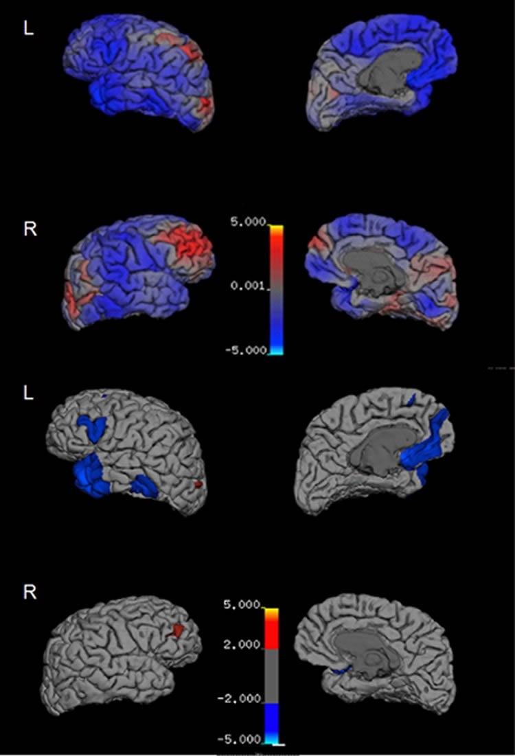
Chronic traumatic encephalopathy (CTE) is a neurodegenerative disorder most commonly associated with repetitive traumatic brain injury (TBI) and characterized by the presence of neurofibrillary tangles of tau protein, known as a tauopathy. Currently, the diagnosis of CTE can only be definitively established postmortem. However, a new positron emission tomography (PET) ligand, [18F]T807/AV1451, may provide the antemortem detection of tau aggregates, and thus various tauopathies, including CTE. Our goal was to examine [18F]T807/AV1451 retention in athletes with neuropsychiatric symptoms associated with a history of multiple concussions. Here we report a 39-year-old retired National Football League player who suffered 22 concussions and manifested progressive neuropsychiatric symptoms. Emotional lability and irritability were the chief complaints. Serial neuropsychological exams revealed a decline in executive functioning, processing speed and fine motor skills. Naming was below average but other cognitive functions were preserved. Structural analysis of longitudinally acquired magenetic resonance imaging scans revealed cortical thinning in the left frontal and lateral temporal areas, as well as volume loss in the basal ganglia. PET with [18F]florbetapir was negative for amyloidosis. The [18F]T807/AV1451 PET showed multifocal areas of retention at the cortical gray matter–white matter junction, a distribution considered pathognomonic for CTE. [18F]T807/AV1451 standard uptake value (SUV) analysis showed increased uptake (SUVrgreater than or equal to1.1) in bilateral cingulate, occipital, and orbitofrontal cortices, and several temporal areas. Although definitive identification of the neuropathological underpinnings basis for [18F]T807/AV1451 retention requires postmortem correlation, our data suggest that [18F]T807/AV1451 tauopathy imaging may be a promising tool to detect and diagnose CTE-related tauopathy in living subjects.
“Cerebral [18 F]T807/AV1451 retention pattern in clinically probable CTE resembles pathognomonic distribution of CTE tauopathy” by D L Dickstein, M Y Pullman, C Fernandez, J A Short, L Kostakoglu, K Knesaurek, L Soleimani, B D Jordan, W A Gordon, K Dams-O’Connor, B N Delman, E Wong, C Y Tang, S T DeKosky, J R Stone, R C Cantu, M Sano, P R Hof and S Gandy in Translational Psychiatry. Published online September 27 2016 doi:10.1038/tp.2016.175


