Age-related EEG patterns correspond with neurophysiological changes associated with development and aging.
Recent Massachusetts General Hospital (MGH) investigations into the neurobiology underlying the effects of general anesthesia have begun to reveal the ways different anesthetic agents alter specific aspects of the brain’s electrical signals, reflected by EEG (electroencephalogram) signatures. While those studies have provided information that may lead to improved techniques for monitoring the consciousness of patients receiving general anesthesia, until now they have been conducted in relatively young adult patients. Now a series of papers from MGH researchers is detailing the differences in the way common anesthetics affect the brains of older patients and children, findings that could lead to ways of improving monitoring technology and the safety of general anesthesia for such patients.
“Anesthesiologists know well that the management of patients age 60 or older requires different approaches than for younger patients,” says Emery Brown, MD, PhD, of the MGH Department of Anesthesia, Critical Care and Pain Medicine. “The doses required to achieve the same anesthetic state in older patients can be as little as half what is needed for younger patients. Explanations for that difference have focused on age-related declines in cardiovascular, respiratory, liver and kidney function, but the primary sites of anesthetic effects are the brain and central nervous system.”
Patrick Purdon, PhD, also of the MGH Department of Anesthesia, Critical Care and Pain Medicine, adds, “We know even less about how anesthetic drugs influence brain activity in children, and the current standard of care for assessing the brain state of children under anesthesia calls only for monitoring vital signs like heart rate and blood pressure. This lack of knowledge is especially troubling, given recent studies suggesting an association between early childhood surgery requiring general anesthesia and later cognitive problems.”
Brown and Purdon lead an MGH research team investigating the neural mechanisms of general anesthesia that in recent years has identified EEG signatures indicating when patients lose and regain consciousness and the EEG patterns – called oscillations – produced by specific drugs while patients are unconscious. In young adults, anesthesia-induced unconsciousness is associated with medium frequency (around 10 Hz) EEG oscillations called frontal alpha waves that are highly synchronized between the cerebral cortex and thalamus, a pattern that is believed to block communication between those brain structures.
Two papers from the MGH team recently published in the British Journal of Anaesthesia are the first to take a detailed look at anesthesia-induced brain changes in older patients. Purdon and Brown are co-corresponding authors of one study that analyzed detailed EEG recordings of 155 patients aged 18 to 90 receiving either propofol or sevoflurane. That study found that the EEG oscillations of older patients were two to three times smaller than those of younger adults with reduced occurrence of frontal alpha waves. The synchronization between the cortex and thalamus occurred at slightly lower frequencies in older patients, who were more likely than younger patients to experience a state called burst suppression that reflects profoundly deep anesthesia at lower doses. The other BJA study, led by MGH anesthesiologist Ken Solt, MD, found that older animals took two to five times longer than younger animals to recover from equal anesthetic doses and observed similar age-related differences in EEG patterns as seen in the patients.

Another study appearing in the same issue of BJA – co-corresponding authors Purdon and Oluwaseun Akeju, MD, MGH Anesthesia, Critical Care and Pain Medicine – analyzed EEG patterns of 54 patients ranging from infancy through age 28 during anesthesia with sevoflurane. They found that anesthesia-induced EEG signals tripled in power from infancy until around age 6 and then dropped off to the typical young adult level at around age 20. Frontal alpha waves were not observed in children under the age of 1, suggesting that the brain circuits required for cortical/thalamic synchronization had not yet developed. Purdon and Brown were also co-authors of an eLife study led by Boston Children’s Hospital investigators Laura Cornelissen, PhD, and Charles Berde, MD, PhD, that detailed the EEG activity of infants 6 months and younger, showing how their patterns evolved toward those more typical of adults over just a few months.
“It appears as though the structure of anesthesia-induced brain dynamics mirrors brain development in children, with different brain wave patterns ‘turning on’ at ages that coincide with known developmental milestones,” says Purdon. “In older patients we see a similar effect but in reverse, with certain brain waves decaying in a manner consistent with brain aging. It’s been known that commercially available EEG-based anesthesia monitors were developed for young adults, and while they are limited for that population – reducing brain activity to a single number – they are even more inaccurate for children and the elderly. These studies illustrate why this is the case and suggest a new, age-specific monitoring paradigm that – along with monitors that track a broader range of EEG signals – could help avoid both anesthesia-induced neurotoxicity in children and post-operative delirium and cognitive dysfunction in elderly patients.”
Brown adds, “Understanding how the brain’s responses to anesthesia change with age allows us to provide personalized, patient-specific strategies for monitoring the brain and dosing the anesthetics, thereby moving us closer to side-effect free anesthesia care.”
Purdon is an assistant professor of Anæsthesia at Harvard Medical School, and Brown is the Warren M. Zapol Professor of Anæsthesia at HMS and the Taplin Professor of Medical Engineering at MIT.
Funding: Support for the MGH-led studies includes several grants from the National Institutes of Health and the Foundation of Anesthesia Education and Research.
Source: Terri Ogan – Massachusetts General Hospital
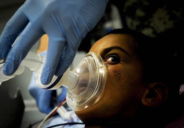
Image Credit: The image is credited to ISAF Headquarters Public Affairs Office and is licensed CC BY 2.0
Original Research: Abstract for “The Ageing Brain: Age-dependent changes in the electroencephalogram during propofol and sevoflurane general anaesthesia” by P. L. Purdon, K. J. Pavone, O. Akeju, A. C. Smith, A. L. Sampson, J. Lee, D. W. Zhou, K. Solt, and E. N. Brown in British Journal of Anaesthesia. Published online April 19 2015 doi:10.1093/bja/aev213
Abstract for “Age-dependency of sevoflurane-induced electroencephalogram dynamics in children” by O. Akeju, K. J. Pavone, J. A. Thum, P. G. Firth, M. B. Westover, M. Puglia, E. S. Shank, E. N. Brown, and P. L. Purdon in British Journal of Anaesthesia. Published online March 27 2015 doi:10.1093/bja/aev114
Abstract
The Ageing Brain: Age-dependent changes in the electroencephalogram during propofol and sevoflurane general anaesthesia
Background Anaesthetic drugs act at sites within the brain that undergo profound changes during typical ageing. We postulated that anaesthesia-induced brain dynamics observed in the EEG change with age.
Methods We analysed the EEG in 155 patients aged 18–90 yr who received propofol (n=60) or sevoflurane (n=95) as the primary anaesthetic. The EEG spectrum and coherence were estimated throughout a 2 min period of stable anaesthetic maintenance. Age-related effects were characterized by analysing power and coherence as a function of age using linear regression and by comparing the power spectrum and coherence in young (18- to 38-yr-old) and elderly (70- to 90-yr-old) patients.
Results Power across all frequency bands decreased significantly with age for both propofol and sevoflurane; elderly patients showed EEG oscillations ∼2- to 3-fold smaller in amplitude than younger adults. The qualitative form of the EEG appeared similar regardless of age, showing prominent alpha (8–12 Hz) and slow (0.1–1 Hz) oscillations. However, alpha band dynamics showed specific age-related changes. In elderly compared with young patients, alpha power decreased more than slow power, and alpha coherence and peak frequency were significantly lower. Older patients were more likely to experience burst suppression.
Conclusions These profound age-related changes in the EEG are consistent with known neurobiological and neuroanatomical changes that occur during typical ageing. Commercial EEG-based depth-of-anaesthesia indices do not account for age and are therefore likely to be inaccurate in elderly patients. In contrast, monitoring the unprocessed EEG and its spectrogram can account for age and individual patient characteristics.
“The Ageing Brain: Age-dependent changes in the electroencephalogram during propofol and sevoflurane general anaesthesia” by P. L. Purdon, K. J. Pavone, O. Akeju, A. C. Smith, A. L. Sampson, J. Lee, D. W. Zhou, K. Solt, and E. N. Brown in British Journal of Anaesthesia. Published online April 19 2015 doi:10.1093/bja/aev213
Abstract
Age-dependency of sevoflurane-induced electroencephalogram dynamics in children
Background General anaesthesia induces highly structured oscillations in the electroencephalogram (EEG) in adults, but the anaesthesia-induced EEG in paediatric patients is less understood. Neural circuits undergo structural and functional transformations during development that might be reflected in anaesthesia-induced EEG oscillations. We therefore investigated age-related changes in the EEG during sevoflurane general anaesthesia in paediatric patients.
Methods We analysed the EEG recorded during routine care of patients between 0 and 28 yr of age (n=54), using power spectral and coherence methods. The power spectrum quantifies the energy in the EEG at each frequency, while the coherence measures the frequency-dependent correlation or synchronization between EEG signals at different scalp locations. We characterized the EEG as a function of age and within 5 age groups: <1 yr old (n=4), 1–6 yr old (n=12), >6–14 yr old (n=14), >14–21 yr old (n=11), >21–28 yr old (n=13).
Results EEG power significantly increased from infancy through ∼6 yr, subsequently declining to a plateau at approximately 21 yr. Alpha (8–13 Hz) coherence, a prominent EEG feature associated with sevoflurane-induced unconsciousness in adults, is absent in patients <1 yr. Conclusions Sevoflurane-induced EEG dynamics in children vary significantly as a function of age. These age-related dynamics likely reflect ongoing development within brain circuits that are modulated by sevoflurane. These readily observed paediatric-specific EEG signatures could be used to improve brain state monitoring in children receiving general anaesthesia.
“Age-dependency of sevoflurane-induced electroencephalogram dynamics in children” by O. Akeju, K. J. Pavone, J. A. Thum, P. G. Firth, M. B. Westover, M. Puglia, E. S. Shank, E. N. Brown, and P. L. Purdon in British Journal of Anaesthesia. Published online March 27 2015 doi:10.1093/bja/aev114


