Summary: Researchers demonstrate how a single injection of fibroblast growth factor 1 (FGF1) can restore blood sugar levels to normal for extended periods in rodent models of type 2 diabetes. Studies show how FGF1 affects specific neurons and perineuronal nets to help restore blood sugar levels to normal, thus sending diabetes into remission.
Source: UW Health
In rodents with type 2 diabetes, a single surgical injection of a protein called fibroblast growth factor 1 can restore blood sugar levels to normal for weeks or months. Yet how this growth factor acts in the brain to generate this lasting benefit has been poorly understood.
Clarifying how this occurs might lead to more effective diabetes treatments that tap into the brain’s inherent potential to ameliorate the condition.
“Until recently, the brain’s ability to normalize elevated blood sugar levels in diabetic animals was unrecognized,” said Dr. Michael Schwartz, professor of medicine at the University of Washington School of Medicine and co-director of the UW Medicine Diabetes Institute.
“By interrogating cellular and molecular responses induced in the hypothalamus by a brain peptide called fibroblast growth factor 1, our international teams’ latest findings chart a path towards a more complete understanding how this effect is achieved.
“These insights,” he said, “may one day inform therapeutic strategies for inducing sustained diabetes remission, rather than simply lowering blood sugar levels on a day-to-day basis as current treatments do.”
Type 2 diabetes affects 10% of the U.S. population. It is closely tied to obesity and causes serious health problems including heart disease, vision loss, kidney failure, dementia, difficult-to-cure infections, and nerve damage. It also increases the risk of needing amputations. Control of blood sugar levels can prevent these problems, but is often hard to achieve and becomes an ongoing struggle for many patients.
In two companion papers in the Sept. 7 editions of Nature Communications and Nature Metabolism, international teams of researchers describe the intricate biology of the brain’s response to fibroblast growth factor 1. The first team describes robust cellular responses that appear to safeguard brain-signaling pathways critical to keeping blood sugar in check.
A second team, containing some of the same researchers, made discoveries about extracellular matrix assemblies called “perineuronal nets” that enmesh groups of neurons involved in blood sugar control. The investigators learned that fibroblast growth factor 1 repairs perineuronal nets that have been damaged by diabetes. This response is required for diabetes remission to be sustained.
Dr. Tunes Pers, of the Novo Nordisk Foundation Center for Basic Metabolic Research, University of Copenhagen in Denmark, and diabetes and obesity researcher Dr. Michael Schwartz at UW Medicine in Seattle were senior authors of the Nature Communications report. The lead authors from their labs were Dr. Marie Bentsen and Dr. Dylan Rausch.
The international team of scientists that they assembled began by detailing changes of gene expression induced by fibroblast growth factor 1 treatment across diverse brain cell types located in the hypothalamus. This small region of the brain regulates many body functions, including levels of blood sugar, hunger, food intake, and energy use and storage.
The scientists found that glial cells, which not only provide structural support but also help to organize and regulate neurocircuit activity, responded more intensely than did neurons, brain cells known for electrical transmission of information.
The researchers also observed enhanced interactions between astrocytes and a subset of neurons that make agouti-related protein (called Agrp neurons). Astrocytes are abundant, star-shaped glial cells that nourish neurons and support their electrical transmissions. Agrp neurons are essential components of the melanocortin signaling system, a brain circuit crucial to control of feeding, body weight and blood sugar.
Excessive activation of Agrp neurons is known to dampen melanocortin signaling. This effect has been linked to diabetes development in people and rodents. The researchers noted that prohibiting melancortin signaling after fibroblast growth factor 1 injection into the brain prevents sustained diabetes remission.
Among other cell types that responded robustly to fibroblast growth factor 1 are tanycytes, elongated, nutrient-sensing glial cells found only in the hypothalamus. Their contributions to normalizing of glucose levels require additional study.
The paper published in Nature Metabolism looked at structures that the scientists called “the previously unrecognized participants” in the mechanism behind fibroblast growth factor 1’s ability to induce diabetes remission.
These are the perineuronal nets that enmesh blood sugar-regulating neurons in the hypothalamus, including Agrp neurons. The lead author of this paper is Kim Alonge, acting instructor in medicine at the UW School of Medicine. The senior author is Michael Schwartz.
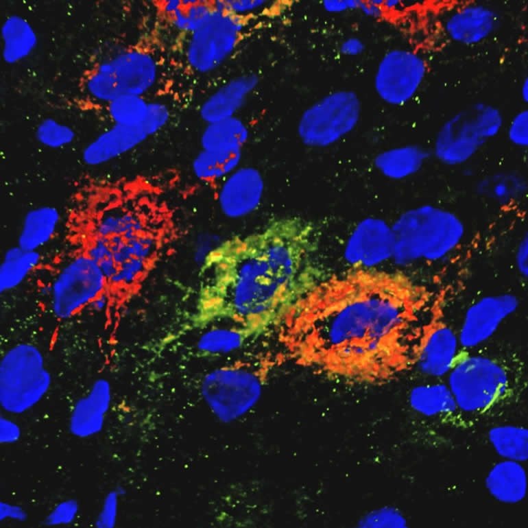
Perineuronal nets promote neurocircuit stability by enmeshing neurons and girding the connections between them. The researchers wanted to know if obesity-related diabetes is associated with structural changes in these perineuronal nets, and whether those could be treated.
The research team noted that in the Zucker Diabetes Fatty rat model of type 2 diabetes, these nets are scarce in the hypothalamus compared to rats with normal blood sugar levels, Yet in other parts of the brain the nets are normal.
This loss of perineuronal nets was rapidly reversed following a single injection of fibroblast growth factor 1 into the brain. The ability of fibroblast growth factor 1 to ameliorate diabetes was hampered by removing the nets through enzymatic digestion. In contrast, intact perineuronal nets are not required for fibroblast growth factor 1 to affect food intake.
These finding identify perineuronal nets as key targets for sustained diabetes remission induced by the action of fibroblast growth factor 1. The researchers speculate that perhaps these nets help to constrain the activity of Agrp neurons and thereby pump up melanocortin signaling.
The researchers plan to continue to try to bridge the gap between the cellular (and extracellular) responses to fibroblast growth factor 1 and the normalization of blood sugar levels. This, they hope, may ultimately uncover novel strategies for achieving sustained diabetes remission in patients.
Funding: The work reported in Nature Metabolism was supported by grants from the National Institute of Diabetes and Digestive and Kidney Disease : P30DK017047 F32DK122662,, R01DK101997, R01DK089056, R01DK083042, and K08DK114474, the National Institute of General Medical Sciences R01GM127579, the National Institute on Aging T32AG052354, the National Institute of Neurological Disorders and Stroke Neurosurgeon Research Career Development Program Award K12NS080223, the Department of Defense Peer Reviewed Medical Research Program W81XWH-20-1-0250 and the Barrow Neurological Foundation18-0025-30-05, the National Institutes of Health Nutrition Obesity Research Center P30DK035816), the UW Royalty Research Fund, Novo Nordisk CMS-431104, Novo Nordisk Foundation, and the Novo Nordisk Foundation Center for Basic Metabolic Research NNF10CC1016515.
The work reported in Nature Communications was supported by grants from the National Institute of Diabetes and Digestive and Kidney Diseases DK-089056 , DK-083042 DK-035816, and DK-101997 and DK-114474, the National Institute of Diabetes and Digestive Kidney Diseases-funded Nutrition Obesity Research Center DK-035816 and Diabetes Research Center DK-017047; Nutrition, Obesity and Atherosclerosis Training Grant T32 HL007028 , Diabetes, Obesity and Metabolism Training Grant T32 DK007247, National Heart, Lung, and Blood Institute Training Grant T32-HL-007312 and Royalty Research Fund at the University of Washington, and the Prometeo Grant for Excellence Research Groups PROMETEO/2019/075, Novo Nordisk Foundation Center for Basic Metabolic Research NNF18CC0034900, Novo Nordisk Foundation 16OC0021496 and NNF17OC0024328, Lundbeck Foundation R19020143904 and R231-2016-3031, and Novo Nordisk CMS-431104.
About this Psychology research article
Source:
UW Health
Contacts:
Leila Gray – UW Health
Image Source:
The image is credited to Kimberly Alonge/Schwartz Lab/UW Medicine Diabetes Research Institute.
Original Research: Open access
“Transcriptomic analysis links diverse hypothalamic cell types to fibroblast growth factor 1-induced sustained diabetes remission” by Marie A. Bentsen, Dylan M. Rausch, Zaman Mirzadeh, Kenjiro Muta, Jarrad M. Scarlett, Jenny M. Brown, Vicente Herranz-Pérez, Arian F. Baquero, Jonatan Thompson, Kimberly M. Alonge, Chelsea L. Faber, Karl J. Kaiyala, Camdin Bennett, Charles Pyke, Cecilia Ratner, Kristoffer L. Egerod, Birgitte Holst, Thomas H. Meek, Burak Kutlu, Yu Zhang, Thomas Sparso, Kevin L. Grove, Gregory J. Morton, Birgitte R. Kornum, José-Manuel García-Verdugo, Anna Secher, Rasmus Jorgensen, Michael W. Schwartz & Tune H. Pers. Nature Communications.
Closed access
“Hypothalamic perineuronal net assembly is required for sustained diabetes remission induced by fibroblast growth factor 1 in rats” by Kimberly M. Alonge, Zaman Mirzadeh, Jarrad M. Scarlett, Aric F. Logsdon, Jenny M. Brown, Elaine Cabrales, Christina K. Chan, Karl J. Kaiyala, Marie A. Bentsen, William A. Banks, Miklos Guttman, Thomas N. Wight, Gregory J. Morton & Michael W. Schwartz. Nature Metabolism.
Abstract
Transcriptomic analysis links diverse hypothalamic cell types to fibroblast growth factor 1-induced sustained diabetes remission
In rodent models of type 2 diabetes (T2D), sustained remission of hyperglycemia can be induced by a single intracerebroventricular (icv) injection of fibroblast growth factor 1 (FGF1), and the mediobasal hypothalamus (MBH) was recently implicated as the brain area responsible for this effect. To better understand the cellular response to FGF1 in the MBH, we sequenced >79,000 single-cell transcriptomes from the hypothalamus of diabetic Lepob/ob mice obtained on Days 1 and 5 after icv injection of either FGF1 or vehicle. A wide range of transcriptional responses to FGF1 was observed across diverse hypothalamic cell types, with glial cell types responding much more robustly than neurons at both time points. Tanycytes and ependymal cells were the most FGF1-responsive cell type at Day 1, but astrocytes and oligodendrocyte lineage cells subsequently became more responsive. Based on histochemical and ultrastructural evidence of enhanced cell-cell interactions between astrocytes and Agrp neurons (key components of the melanocortin system), we performed a series of studies showing that intact melanocortin signaling is required for the sustained antidiabetic action of FGF1. These data collectively suggest that hypothalamic glial cells are leading targets for the effects of FGF1 and that sustained diabetes remission is dependent on intact melanocortin signaling.
Abstract
Hypothalamic perineuronal net assembly is required for sustained diabetes remission induced by fibroblast growth factor 1 in rats
We recently showed that perineuronal nets (PNNs) enmesh glucoregulatory neurons in the arcuate nucleus (Arc) of the mediobasal hypothalamus (MBH)1, but whether these PNNs play a role in either the pathogenesis of type 2 diabetes (T2D) or its treatment remains unclear. Here we show that PNN abundance within the Arc is markedly reduced in the Zucker diabetic fatty (ZDF) rat model of T2D, compared with normoglycaemic rats, correlating with altered PNN-associated sulfation patterns of chondroitin sulfate glycosaminoglycans in the MBH. Each of these PNN-associated changes is reversed following a single intracerebroventricular (icv) injection of fibroblast growth factor 1 (FGF1) at a dose that induces sustained diabetes remission in male ZDF rats. Combined with previous work localizing this FGF1 effect to the Arc area2,3,4, our finding that enzymatic digestion of Arc PNNs markedly shortens the duration of diabetes remission following icv FGF1 injection in these animals identifies these extracellular matrix structures as previously unrecognized participants in the mechanism underlying diabetes remission induced by the central action of FGF1.