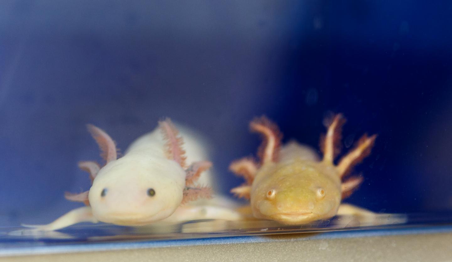
Summary: Axolotl salamander genes that allow the neural tube and nerve fibers to regenerate after spinal cord damage have been identified. These genes are also found in humans, but are activated differently.
Source: Marine Biological Laboratory
Researchers are one step closer to solving the mystery of why some vertebrates can regenerate their spinal cords while others, including humans, create scar tissue after spinal cord injury, leading to lifelong damage.
Scientists at the Marine Biological Laboratory (MBL) have identified gene “partners” in the axolotl salamander that, when activated, allow the neural tube and associated nerve fibers to functionally regenerate after severe spinal cord damage. Interestingly, these genes are also present in humans, though they are activated in a different manner. Their results are published this week in Nature Communications Biology.
“[Axolotls are] the champions of regeneration in that they can regenerate multiple body parts. For example, if you make a lesion in the spinal cord, they can fully regenerate it and gain back both motor and sensory control,” says Karen Echeverri, associate scientist in the Eugene Bell Center for Regenerative Biology and Tissue Engineering. “We wanted to understand what is different at a molecular level that drives them towards this pro-regenerative response instead of forming scar tissue.”
Echeverri’s prior research had shown that, in both axolotls and humans, the c-Fos gene is up-regulated in the glial cells of the nervous system after spinal cord injury. She also knew that c-Fos cannot act alone.
“It’s what we call an obligate heterodimer, so it has to have a partner in life,” says Echeverri. “c-Fos has a different partner in axolotl than it has in humans and this seems to drive a completely different response to injury.”

In human injury response, c-Fos is paired with the gene c-Jun. In axolotls, however, Echeverri and her team determined that c-Fos is activated with the gene JunB. This difference in gene activation was traced to the actions of microRNAs, which regulate gene expression.
By modifying gene expression by the axolotls’ microRNA, they were able to force the human pairing of c-Fos with c-Jun. The salamanders with the human pairings were unable to regain a functioning spinal cord after injury, instead forming the scar tissue that occurs in human injury repair. Follow-up studies will investigate if the reverse is true in human cells.
“The genes involved in regeneration in axolotl are highly conserved between humans and axolotls, and it doesn’t appear so far that axolotls have regeneration-specific genes,” says Echeverri. “It’s all about who you partner with directly after injury, and how that drives you toward either regeneration or forming scar tissue. It’s kind of like in life, who you partner with can have a really positive or negative effect.”
Understanding the axolotl spinal cord regeneration and its differences from–and, more interestingly, similarities to–the human process could help researchers and eventually doctors improve treatment for severe human spinal cord injuries.
“That has a huge translation potential not just for spinal cord injury in humans, but also for a lot of neurodegenerative diseases,” Echeverri says.
“MBL has a history of making basic discoveries in research organisms that the general person on the street may not think have any relevance to human health,” she says. “But major discoveries have been made from studying animals like axolotls or jelly fish, and here at MBL we have that freedom to work on those really basic questions.”
Written by Stephanie McPherson
Source: Marine Biological Laboratory
Media Contact: Diana Kenney – Marine Biological Laboratory
Publisher: Scientists Identify Gene Partnerships that Promote Spinal Cord Regeneration organized by Neuroscience News.
Image Source: Axolotis image is credited to Dee Sullivan.
Original Research: Abstract for ” AP-1cFos/JunB/miR-200a regulate the pro-regenerative glial cell response during axolotl spinal cord regeneration” by Keith Z. Sabin, Peng Jiang, Micah D. Gearhart, Ron Stewart & Karen Echeverri in Nature Communications Biology. Published March 6, 2019
doi: 10.1038/s42003-019-0335-4
Funding: National Institutes of Health
Abstract
AP-1cFos/JunB/miR-200a regulate the pro-regenerative glial cell response during axolotl spinal cord regeneration
Salamanders have the remarkable ability to functionally regenerate after spinal cord transection. In response to injury, GFAP+ glial cells in the axolotl spinal cord proliferate and migrate to replace the missing neural tube and create a permissive environment for axon regeneration. Molecular pathways that regulate the pro-regenerative axolotl glial cell response are poorly understood. Here we show axolotl glial cells up-regulate AP-1cFos/JunB after injury, which promotes a pro-regenerative glial cell response. Injury induced upregulation of miR-200a in glial cells supresses c-Jun expression in these cells. Inhibition of miR-200a during regeneration causes defects in axonal regrowth and transcriptomic analysis revealed that miR-200a inhibition leads to differential regulation of genes involved with reactive gliosis, the glial scar, extracellular matrix remodeling and axon guidance. This work identifies a unique role for miR-200a in inhibiting reactive gliosis in axolotl glial cells during spinal cord regeneration.


