Summary: A new study reports a gene mutation linked to autism also plays a critical role when it comes to the formation and maturation of synapses.
Source: MIT.
Loss of Shank gene prevents neuronal synapses from properly maturing.
A new study from MIT neuroscientists reveals that a gene mutation associated with autism plays a critical role in the formation and maturation of synapses — the connections that allow neurons to communicate with each other.
Many genetic variants have been linked to autism, but only a handful are potent enough to induce the disorder on their own. Among these variants, mutations in a gene called Shank3 are among the most common, occurring in about 0.5 percent of people with autism.
Scientists know that Shank3 helps cells respond to input from other neurons, but because there are two other Shank proteins, and all three can fill in for each other in certain ways, it has been difficult to determine exactly what the Shank proteins are doing.
“It’s clearly regulating something in the neuron that’s receiving a synaptic signal, but some people find one role and some people find another,” says Troy Littleton, a professor in the departments of Biology and of Brain and Cognitive Sciences at MIT, a member of MIT’s Picower Institute for Learning and Memory, and the senior author of the study. “There’s a lot of debate over what it really does at synapses.”
Key to the study is that fruit flies, which Littleton’s lab uses to study synapses, have only one version of the Shank gene. By knocking out that gene, the researchers eliminated all Shank protein from the flies.
“This is the first animal where we have completely removed all Shank family proteins,” says Kathryn Harris, a Picower Institute research scientist and lead author of the paper, which appears in the May 25 issue of the Journal of Neuroscience.
Synaptic organization
Scientists already knew that the Shank proteins are scaffold proteins, meaning that they help to organize the hundreds of other proteins found in the synapse of a postsynaptic cell — a cell that receives signals from a presynaptic cell. These proteins help to coordinate the cell’s response to the incoming signal.
“Shank is essentially a hub for signaling,” Harris says. “It brings in a lot of other partners and plays an organizational role at the postsynaptic membrane.”
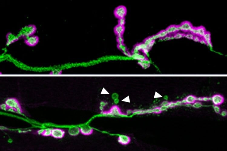
In fruit flies lacking the Shank protein, the researchers found two dramatic effects. First, the postsynaptic cells had many fewer boutons, which are the sites where neurotransmitter release occurs.
Second, many of the boutons that did form were not properly developed; that is, they were not surrounded by all of the postsynaptic proteins normally found there, which are required to respond to synaptic signals.
The researchers are now studying how this reduction in functional synapses affects the brain. Littleton suspects that the development of neural circuits could be impaired, which, if the same holds true in humans, may help explain some of the symptoms seen in autistic people.
“During critical windows of social and language learning, we reshape our connections to drive connectivity patterns that respond to rewards and language and social interactions,” he says. “If Shank is doing similar things in the mammalian brain, one could imagine potentially having those circuits form relatively normally early on, but if they fail to properly mature and form the proper number of connections, that could lead to a variety of behavioral defects.”
Pinpointing an exact link to autism symptoms would be difficult to do in fruit fly studies, however.
“Although the core molecular machines that allow neurons to communicate are highly conserved between fruit flies and humans, the anatomy of the various circuits that are formed during evolution are quite different,” Littleton says. “It’s hard to jump from a synaptic defect in the fly to an autism-like phenotype because the circuits are so different.”
An unexpected link
The researchers also showed, for the first time, that loss of Shank affects a well-known set of proteins that comprise the Wnt (also known as Wingless) signaling pathway. When a Wnt protein binds to a receptor on the cell, it initiates a series of interactions that influence which genes are turned on. This, in turn, contributes to many cell processes including embryonic development, tissue regeneration, and tumor formation.
When Shank is missing from fruit flies, Wnt signaling is disrupted because the receptor that normally binds to Wnt fails to be internalized by the cell. Normally, a small segment of the activated receptor moves to the cell nucleus and influences the transcription of genes that promote maturation of synapses. Without Shank, Wnt signaling is impaired and the synapses do not fully mature.
“The Shank protein and the Wnt protein family are thought to be involved in autism independently, but the fact that this study discovered that Wnt and Shank are interacting brings the story into better focus,” says Bryan Stewart, a professor of cell and systems biology at the University of Toronto, who was not involved in the research. “Now we can look and see if those interactions between Wnt and Shank are potentially responsible for their role in autism.”
The finding raises the possibility of treating autism with drugs that promote Wnt signaling, if the same connection is found in humans.
“Because the link to Wnt signaling is new and hasn’t been picked up in mammalian studies, we really hope that that can inspire people to look for a connection to Wnt signaling in mammalian models, and maybe that can offer another avenue for how loss of Shank could be counteracted,” Harris says.
Funding: The research was funded by the National Institutes of Health and the Simons Center for the Social Brain at MIT.
Source: Anne Trafton – MIT
Image Source: This NeuroscienceNews.com image is credited to the researchers.
Original Research: The study will appear in Journal of Neuroscience.
[cbtabs][cbtab title=”MLA”]MIT. “Illuminating the Role of Autism Linked Shank Gene.” NeuroscienceNews. NeuroscienceNews, 25 May 2016.
<https://neurosciencenews.com/autism-shank3-synapses-4297/>.[/cbtab][cbtab title=”APA”]MIT. (2016, May 25). Illuminating the Role of Autism Linked Shank Gene. NeuroscienceNews. Retrieved May 25, 2016 from https://neurosciencenews.com/autism-shank3-synapses-4297/[/cbtab][cbtab title=”Chicago”]MIT. “Illuminating the Role of Autism Linked Shank Gene.” https://neurosciencenews.com/autism-shank3-synapses-4297/ (accessed May 25, 2016).[/cbtab][/cbtabs]