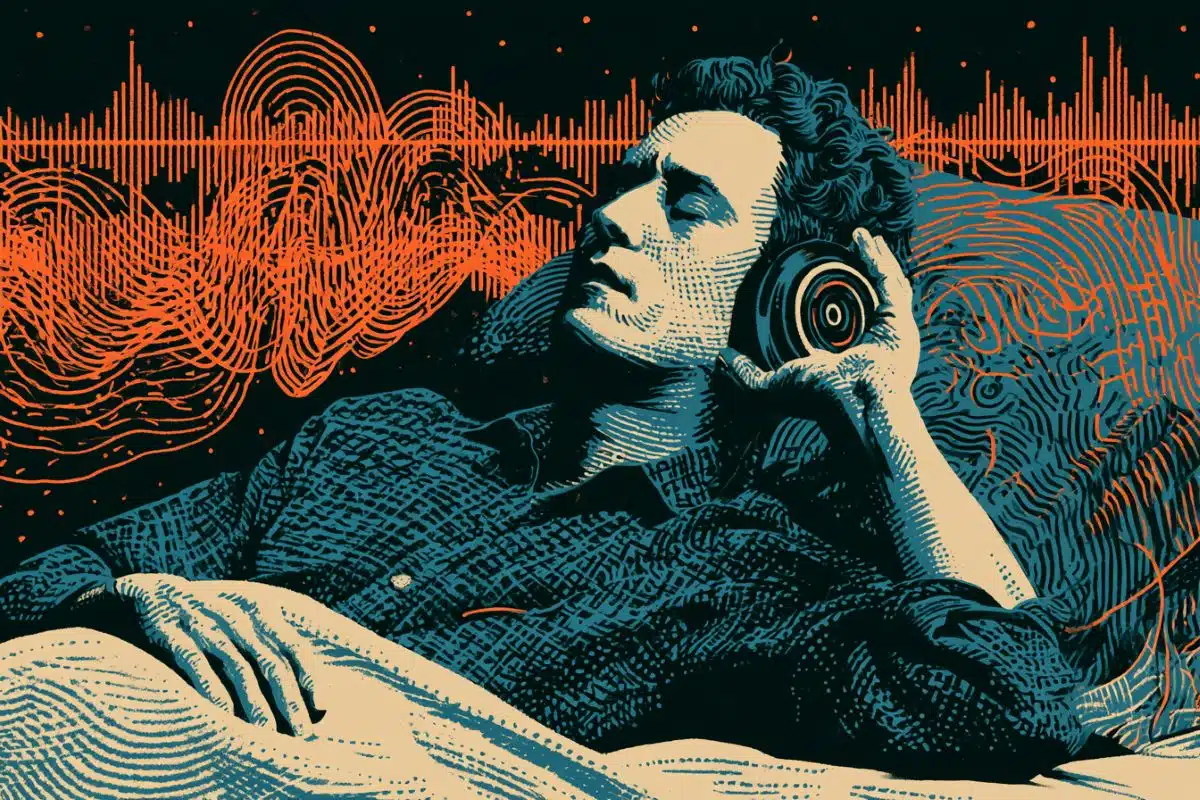
Summary: Virtual reality is helping researchers uncover some of the secrets of anxiety. Using VR, study participants were able to distinguish between safe and dangerous environments in a game. However, brain scans of those with anxiety showed increased activity in the insula and dorsomedial prefrontal cortex while in a safe zone, indicating their brains were associating the safe environment with threat or danger.
Source: Rutgers University
Imagine you are in a meadow picking flowers. You know that some flowers are safe, while others have a bee inside that will sting you. How would you react to this environment and, more importantly, how would your brain react?
This is the scene in a virtual-reality environment used by researchers to understand the impact anxiety has on the brain and how brain regions interact with one another to shape behavior.
“These findings tell us that anxiety disorders might be more than a lack of awareness of the environment or ignorance of safety, but rather that individuals suffering from an anxiety disorder cannot control their feelings and behavior even if they wanted to,” said Benjamin Suarez-Jimenez, Ph.D., assistant professor in the Del Monte Institute for Neuroscience at the University of Rochester and first author of the study published in Communications Biology.
“The patients with an anxiety disorder could rationally say – I’m in a safe space – but we found their brain was behaving as if it was not.”
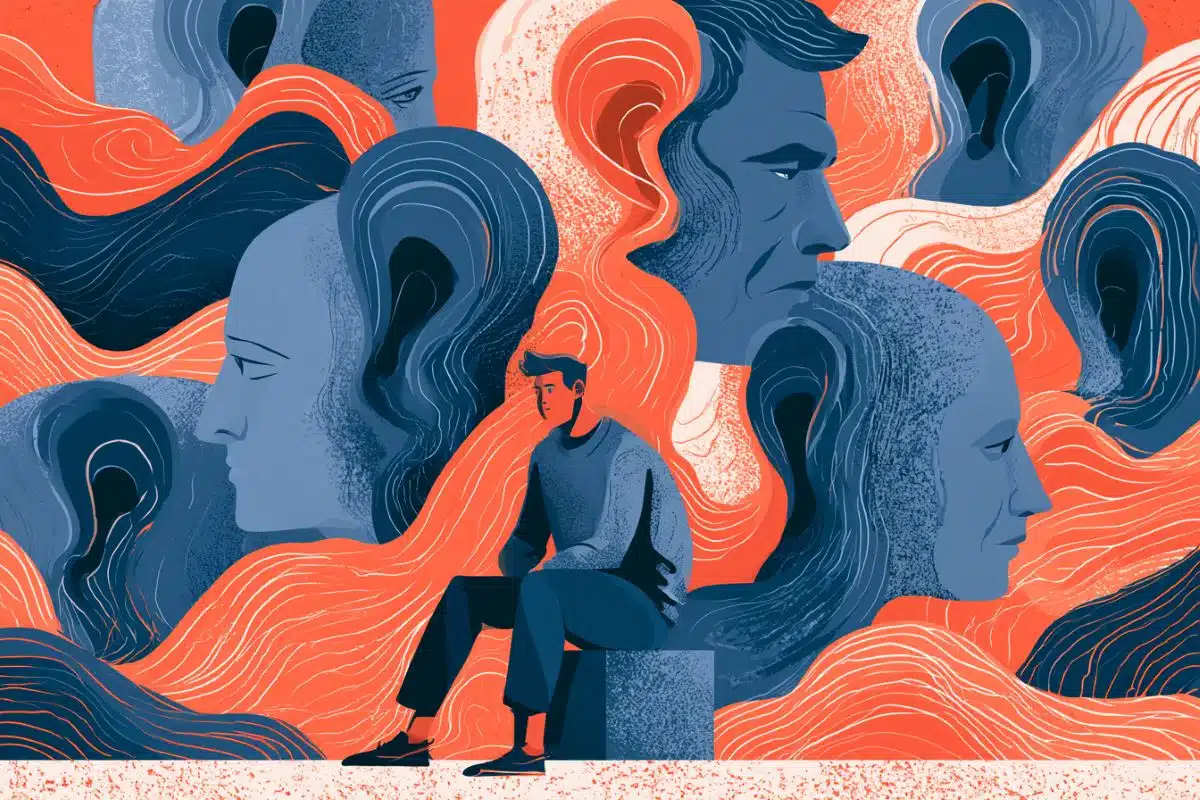
Watching anxiety in the brain
Using fMRI, the researchers observed the brain activity of volunteers with general and social anxiety as they navigated a virtual reality game of picking flowers. Half of the meadow had flowers without bees, the other half had flowers with bees that would sting them – as simulated by a mild electrical stimulation to the hand.
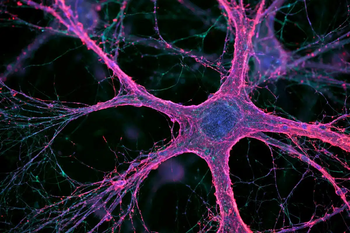
Researchers found all study participants could distinguish between the safe and dangerous areas, however, brain scans revealed volunteers with anxiety had increased insula and dorsomedial prefrontal cortex activation – indicating their brain was associating a known safe area to danger or threat.
“This is the first time we’ve looked at discrimination learning in this way. We know what brain areas to look at, but this is the first time we show this concert of activity in such a complex ‘real-world-like’ environment,” said Suarez-Jimenez. “These findings point towards the need for treatments that focus on helping patients take back control of their body.”
The brain differences were the only differences seen in these patients. For example, sweat responses, a proxy for anxiety, which was also measured, failed to reveal any clear differences.
Suarez-Jimenez’s research
Understanding the neural mechanisms by which the brain learns about the environment is the focus of Suarez-Jimenez’s research, particularly how the brain predicts what is threatening and what is safe. He uses virtual reality environments to investigate neural signatures of anxiety disorders and post-traumatic stress disorder (PTSD). His goal is to understand how people build maps in the brain that are based on experience, and the role of those maps in psychopathologies of stress and anxiety.

Expanding research to other disorders
“For next steps in this recent research, we still need to clarify if what we found in the brain of these patients is also the case in other disorders, such as PTSD. Understanding the differences and similarities across disorders characterized by deficits in behavioral regulation and feelings in safe environments, can help us create better personalized treatment options.”
Monique Ernst, M.D., Ph.D., with the National Institute of Mental Health (NIMH) was senior author on the paper. Co-authors include Nicholas Balderston, Ph.D., Joseph Leshin, Abigail Hsiung, Daniel Pine, M.D., and Christian Grillon, Ph.D., of the NIMH, along with James Bisby, Ph.D., John King, Ph.D., and Neil Burgess, Ph.D., of the University College London.
Funding: The research reported in this press release was supposed by was funded by the NIMH Intramural Research Program of the National Institutes of Health under award numbers MH015144, and MH118428-01, the Medical Research Council, and the Wellcome Trust, United Kingdom. The content is solely the responsibility of the authors and does not necessarily represent the official views of the National Institutes of Health.
About this anxiety research news
Author: Kelsie Smith Hayduk
Source: Rutgers University
Contact: Kelsie Smith Hayduk – Rutgers University
Image: The image is in the public domain
Original Research: Open access.
“Location-dependent threat and associated neural abnormalities in clinical anxiety” by Benjamin Suarez-Jimenez et al. Communications Biology
Abstract
Location-dependent threat and associated neural abnormalities in clinical anxiety
Anxiety disorders are characterized by maladaptive defensive responses to distal or uncertain threats.
Elucidating neural mechanisms of anxiety is essential to understand the development and maintenance of anxiety disorders. In fMRI, patients with pathological anxiety (ANX, n = 23) and healthy controls (HC, n = 28) completed a contextual threat learning paradigm in which they picked flowers in a virtual environment comprising a danger zone in which flowers were paired with shock and a safe zone (no shock). ANX compared with HC showed 1) decreased ventromedial prefrontal cortex and anterior hippocampus activation during the task, particularly in the safe zone, 2) increased insula and dorsomedial prefrontal cortex activation during the task, particularly in the danger zone, and 3) increased amygdala and midbrain/periaqueductal gray activation in the danger zone prior to potential shock delivery.
Findings suggest that ANX engage brain areas differently to modulate context-appropriate emotional responses when learning to discriminate cues within an environment.