

Summary: Study shows tau tangles in the brain may be the driving mechanism of Alzheimer’s pathology.
Source: SNM.
Alzheimer’s is a devastating and incurable disease marked by beta-amyloid and tau protein aggregations in the brain, yet the direct relationship between these proteins and neurodegeneration has remained a mystery. New molecular imaging research is revealing how tau, rather than amyloid-deposition, may be more directly instigating neuronal dysfunction, say presenters at the 2016 Annual Meeting of the Society of Nuclear Medicine and Molecular Imaging (SNMMI).
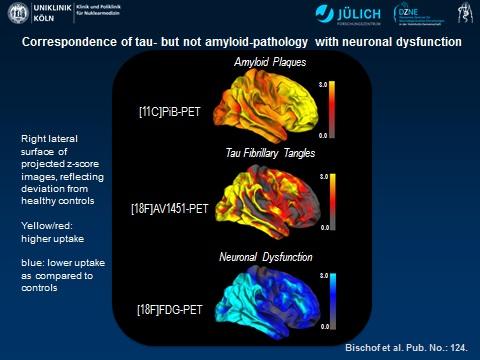
Exhaustive brain research has pieced together how extracellular beta-amyloid plaques and intracellular neurofibrillary tangles of tau proteins are strongly linked to the neurodegenerative pathology of Alzheimer’s disease. However, much of the research conducted to date has been post-mortem, which does little to help researchers understand the early development of disease. A new imaging study in living Alzheimer’s patients is combining three methods of positron emission tomography (PET) to measure the orchestration of amyloid, tau and metabolic activity in the brain. Findings of the study showed a significant correlation between increased tau and decreased metabolic activity in the brain, a clear sign of neurodegeneration.
“Tau-imaging seems to be closely linked to actual onset of neuronal injury, whereas amyloid-imaging may allow us to detect a predisposition to disease many years ahead of the onset of symptoms,” said Alexander Drzezga, MD, from the German Center for Neurodegenerative Diseases at the University Hospital of Cologne in Cologne, Germany.
For this study, 10 subjects with Alzheimer’s underwent PET following the injection of three radiotracers: fluorine-18 fluorodeoxyglucose (F-18 FDG), which images regional metabolic activity; carbon-11 Pittsburgh compound B (C-11 PiB), which has an affinity for amyloid plaques; and F-18 AV-1451, an emerging imaging agent that binds to tau in the brain. Results showed that increased tau was directly associated with hypometabolism (reflecting neuronal dysfunction) in the brain. For amyloid-deposition, no strong association with hypometabolism was found. However, an indirect interactivity between tau and amyloid was observed particularly in the parietal cortex, in that the negative impact of regional tau-deposition on metabolism was stronger in regions with higher amyloid-burden.
“Integrating these molecular imaging tools offers the opportunity to investigate the possible independent and synergistic contribution of these protein pathologies in neurodegeneration in the living brain and, therefore, greatly advance our understanding of the mechanisms of Alzheimer’s disease,” said Drzezga.
Further investigation of these and other factors of neurodegeneration in living dementia patients could one day help clinicians improve diagnostic accuracy and lead to disease-modifying therapies for Alzheimer’s, including new drugs that could potentially target tau in order to slow or stop degenerative effects in the brain. Multimodal imaging approaches like this one could allow more precise staging of neuropathology, even before the irrevocable onset of memory loss experienced by Alzheimer’s patients. Furthermore, improved prediction, prognosis and therapy control/follow up may become feasible.
More than 46 million people are currently living with Alzheimer’s across the world, and that number is expected to rise steeply to 131.5 million by 2050. The global economic cost of the disease is expected to approach $1 trillion in the same period, according to the newest data from Alzheimer’s Disease International.
Source: Laurie Callahan – SNM
Image Source: This NeuroscienceNews.com image is credited to G. Bischof, J. Hammes, T. van Eimeren, A. Drzezga.
Original Research: The findings will be presented at SNMMI’s 63rd Annual Meeting, June 11-15, 2016, San Diego.
[cbtabs][cbtab title=”MLA”]SNM. “PET Imaging Points to Tau Protein as Leading Culprit in Alzheimer’s.” NeuroscienceNews. NeuroscienceNews, 13 June 2016.
<https://neurosciencenews.com/tau-alzheimers-pet-imaging-4459/>.[/cbtab][cbtab title=”SNM”]SNM. (2016, June 13). PET Imaging Points to Tau Protein as Leading Culprit in Alzheimer’s. NeuroscienceNews. Retrieved June 13, 2016 from https://neurosciencenews.com/tau-alzheimers-pet-imaging-4459/[/cbtab][cbtab title=”Chicago”]SNM. “PET Imaging Points to Tau Protein as Leading Culprit in Alzheimer’s.” https://neurosciencenews.com/tau-alzheimers-pet-imaging-4459/ (accessed June 13, 2016).[/cbtab][/cbtabs]


