

Summary: A new study reveals nanoparticles from tattoo ink travels into the body and reaches the lymph nodes. Researchers have collected visual evidence showing lymph nodes tinted with the colors of a person’s tattoos.
Source: ESRF.
The elements that make up the ink in tattoos travel inside the body in micro and nanoparticle forms and reach the lymph nodes according to a study published in Scientific Reports on 12 September by scientists from Germany and the ESRF, the European Synchrotron, Grenoble (France). It is the first time that there is analytical evidence of the transport of various organic and inorganic pigments and toxic element impurities as well as in depth characterization of the pigments ex vivo in tattooed tissues. Two ESRF beamlines have been crucial in this breakthrough.
“When someone wants to get a tattoo, they are often very careful in choosing a parlour where they use sterile needles that haven´t been used previously. No one checks the chemical composition of the colours, but our study shows that maybe they should”, explains Hiram Castillo, one of the authors of the study and scientist at the ESRF.
The reality is that little is known about the potential impurities in the colour mixture applied to the skin. Most tattoo inks contain organic pigments, but also include preservatives and contaminants like nickel, chromium, manganese or cobalt. Besides carbon black, the second most common ingredient used in tattoo inks is titanium dioxide (TiO2), a white pigment usually applied to create certain shades when mixed with colorants. TiO2 is also commonly used in food additives, sun screens, paints. Delayed healing, along with skin elevation and itching, are often associated with white tattoos, and by consequence with the use of TiO2. Scientists from the ESRF, the German Federal Institute for Risk Assessment, Ludwig-Maximilians University, and the Physikalisch-Technische Bundesanstalt have managed to get a very clear picture on the location of titanium dioxide once they get in the tissue. This work was done on the ESRF beamlines or experimental stations, ID21 and ID16B.
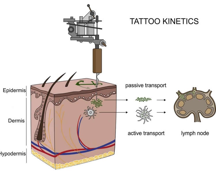
The hazards that potentially derive from tattoos were as yet only investigated by chemical analysis of the inks and their degradation products in vitro. “We already knew that pigments from tattoos would travel to the lymph nodes because of visual evidence: the lymph nodes become tinted with the colour of the tattoo. It is the response of the body to clean the site of entrance of the tattoo. What we didn’t know is that they do it in a nano form, which implies that they may not have the same behaviour as the particles at a micro level. And that is the problem: we don’t know how nanoparticles react”, explains Bernhard Hesse, one of the two first authors of the study and ESRF visiting scientist.
X-ray fluorescence measurements on ID21 allowed the team to locate titanium dioxide at the micro and nano range in the skin and the lymphatic environment. They found a broad range of particles with up to several micrometres in size in human skin but only smaller (nano) particles transported to the lymph nodes. This may lead to the chronic enlargement of the lymph node and lifelong exposure. Scientists also used the technique of Fourier transform infrared spectroscopy to assess biomolecular changes in the tissues in the proximity of the tattoo particles.
Altogether the scientists report strong evidence for both migration and long-term deposition of toxic elements and tattoo pigments as well as for conformational alterations of biomolecules that are sometimes linked to cutaneous inflammation and other adversities upon tattooing.
Then next step for the team is to inspect further samples of patients with adverse effects in their tattoos in order to find links with chemical and structural properties of the pigments used to create these tattoos.
Funding: This study was funded by the German Federal Institute for Risk Assessment.
Source: Delphine Chenevier – ESRF
Image Source: NeuroscienceNews.com image is in the public domain. The diagram is credited to Christian Seim.
Original Research: Full open access research for “Synchrotron-based ν-XRF mapping and μ-FTIR microscopy enable to look into the fate and effects of tattoo pigments in human skin” by Ines Schreiver, Bernhard Hesse, Christian Seim, Hiram Castillo-Michel, Julie Villanova, Peter Laux, Nadine Dreiack, Randolf Penning, Remi Tucoulou, Marine Cotte & Andreas Luch in Scientific Reports. Published online September 12 2017 doi:10.1038/s41598-017-11721-z
[cbtabs][cbtab title=”MLA”]ESRF “Tattoos Are More Than Skin Deep: Ink Can Reach Lymph Nodes.” NeuroscienceNews. NeuroscienceNews, 16 September 2017.
<https://neurosciencenews.com/tattoo-ink-lymph-node-7506/>.[/cbtab][cbtab title=”APA”]ESRF (2017, September 16). Tattoos Are More Than Skin Deep: Ink Can Reach Lymph Nodes. NeuroscienceNews. Retrieved September 16, 2017 from https://neurosciencenews.com/tattoo-ink-lymph-node-7506/[/cbtab][cbtab title=”Chicago”]ESRF “Tattoos Are More Than Skin Deep: Ink Can Reach Lymph Nodes.” https://neurosciencenews.com/tattoo-ink-lymph-node-7506/ (accessed September 16, 2017).[/cbtab][/cbtabs]
Abstract
Synchrotron-based ν-XRF mapping and μ-FTIR microscopy enable to look into the fate and effects of tattoo pigments in human skin
The increasing prevalence of tattoos provoked safety concerns with respect to particle distribution and effects inside the human body. We used skin and lymphatic tissues from human corpses to address local biokinetics by means of synchrotron X-ray fluorescence (XRF) techniques at both the micro (μ) and nano (ν) scale. Additional advanced mass spectrometry-based methodology enabled to demonstrate simultaneous transport of organic pigments, heavy metals and titanium dioxide from skin to regional lymph nodes. Among these compounds, organic pigments displayed the broadest size range with smallest species preferentially reaching the lymph nodes. Using synchrotron μ-FTIR analysis we were also able to detect ultrastructural changes of the tissue adjacent to tattoo particles through altered amide I α-helix to β-sheet protein ratios and elevated lipid contents. Altogether we report strong evidence for both migration and long-term deposition of toxic elements and tattoo pigments as well as for conformational alterations of biomolecules that likely contribute to cutaneous inflammation and other adversities upon tattooing.
“Synchrotron-based ν-XRF mapping and μ-FTIR microscopy enable to look into the fate and effects of tattoo pigments in human skin” by Ines Schreiver, Bernhard Hesse, Christian Seim, Hiram Castillo-Michel, Julie Villanova, Peter Laux, Nadine Dreiack, Randolf Penning, Remi Tucoulou, Marine Cotte & Andreas Luch in Scientific Reports. Published online September 12 2017 doi:10.1038/s41598-017-11721-z


