

Finding provides alternative explanation for the free-radical theory of aging and suggests new role for mitochondria in affecting physiology.
Buck Institute faculty Judith Campisi, PhD, says age researchers need to stop thinking of cellular senescence, now accepted as an important driver of aging, as a single phenotype that stems from genotoxic stress. Research from her lab reveals that cellular senescence, a process whereby cells permanently lose the ability to divide, is also induced by signaling from dysfunctional mitochondria – and that the arrested cells secrete a distinctly different “stew” of biologically active factors in a process unrelated to the damaging free radicals that are created in mitochondria as part of oxygen metabolism. The results are published in Cell Metabolism.
“We don’t yet know how much this process contributes to natural aging,” said Campisi, adding that those studies are currently underway. “But we do think the findings are important in addressing mitochondrial diseases, and those age-related diseases, such as some forms of Parkinson’s, which involve mitochondrial dysfunction.”
The discovery was unexpected and was made by postdoctoral fellow Christopher Wiley, PhD, who (in collaboration with Eric Verdin, PhD, from the Gladstone Institute) was eliminating sirtuins, a class of proteins long linked to longevity, one by one in human cell cultures. “The senescent phenotype only occurred when we eliminated the mitochondrial sirtuins,” said Wiley. In addition, Wiley saw that the senescent cells secreted a different SASP (senescence-associated secretory phenotype) than expected – one that lacks the IL-1-dependent inflammatory arm – a major factor in the SASP originally identified in the Campisi lab in 2008. The authors dubbed this new phenomenon MiDAS – mitochondrial dysfunction-associated senescence.
Along with identifying MiDAS, Wiley also found that mitochondrial dysfunction upset the balance of NAD+ (an enzyme that is a co-factor for sirtuins), which arrested cell growth and prevented the IL-1-associated SASP. “The NAD+ balancing act happens outside the mitochondria in the cytoplasm of the cell,” said Wiley. “This really highlights a signaling role for mitochondria, something understudied in the context of disease. And it identifies a new type of SASP, underscoring the existence of different types of senescence.”
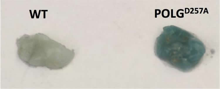
Wiley also identified the MiDAS SASP in mice genetically engineered to develop progeria in response to mitochondrial mutations, which causes rapid aging. The MiDAS SASP suppressed adipogenesis, which plays a vital role in metabolism and the creation of fat cells. He says the work provides a link to lipodystrophy, a medical condition characterized by abnormal or degenerative conditions involving fat tissue, adding that early HIV drugs (which are still being used in third world countries) deplete mitochondrial DNA and that patients receiving the drugs often show a particular loss of subcutaneous fat, especially in their face.
“For any disease that has a mitochondrial component this research adds a potential explanation for the real driver of the dysfunction — and it’s not free radicals, which we ruled out in our study” said Campisi. “Our finding suggest a new role for mitochondria when it comes to affecting physiology.”
Other Buck researchers involved in the study include Michael C. Velarde, Pacome Lecot, Su Liu, Ethan A. Sarnoski, Adam Freund, Sonnet S. Davis, Arvind Ramanathan, and Akos A. Gerencser. Collaborators from the Gladstone Institute include Eric Verdin, Kotaro Shirakawa and Hyung W. Lim.
Funding: The work was funded by NIH grants T32-AG00266, R37-AG009909, K-99-AG041221 and fellowships from the American Federation of Aging Research and the SENS Research Foundation and ExploRA’, PULSE and CROUS from France.
Source: Kris Rebillot – Buck Institute
Image Source: The image is credited to Akos Gerencser, PhD, Buck Institute
Original Research: Abstract for “Mitochondrial Dysfunction Induces Senescence with a Distinct Secretory Phenotype” by Christopher D. Wiley, Michael C. Velarde, Pacome Lecot, Su Liu, Ethan A. Sarnoski, Adam Freund, Kotaro Shirakawa, Hyung W. Lim, Sonnet S. Davis, Arvind Ramanathan, Akos A. Gerencser, Eric Verdin, and Judith Campisi in Cell Metabolism. Published online December 10 2015 doi:10.1016/j.cmet. 2015.11.011
Abstract
Mitochondrial Dysfunction Induces Senescence with a Distinct Secretory Phenotype
Highlights
•Dysfunctional mitochondria cause cell senescence and a distinct secretory phenotype
•This secretory phenotype can influence the differentiation of certain cell types
•An NAD-AMPK-p53 pathway controls the secretory and mitotic arrest phenotypes
•Mice with dysfunctional mitochondria and premature aging accumulate senescent cells
Summary
Cellular senescence permanently arrests cell proliferation, often accompanied by a multi-faceted senescence-associated secretory phenotype (SASP). Loss of mitochondrial function can drive age-related declines in the function of many post-mitotic tissues, but little is known about how mitochondrial dysfunction affects mitotic tissues. We show here that several manipulations that compromise mitochondrial function in proliferating human cells induce a senescence growth arrest with a modified SASP that lacks the IL-1-dependent inflammatory arm. Cells that underwent mitochondrial dysfunction-associated senescence (MiDAS) had lower NAD+/NADH ratios, which caused both the growth arrest and prevented the IL-1-associated SASP through AMPK-mediated p53 activation. Progeroid mice that rapidly accrue mtDNA mutations accumulated senescent cells with a MiDAS SASP in vivo, which suppressed adipogenesis and stimulated keratinocyte differentiation in cell culture. Our data identify a distinct senescence response and provide a mechanism by which mitochondrial dysfunction can drive aging phenotypes.
“Mitochondrial Dysfunction Induces Senescence with a Distinct Secretory Phenotype” by Christopher D. Wiley, Michael C. Velarde, Pacome Lecot, Su Liu, Ethan A. Sarnoski, Adam Freund, Kotaro Shirakawa, Hyung W. Lim, Sonnet S. Davis, Arvind Ramanathan, Akos A. Gerencser, Eric Verdin, and Judith Campisi in Cell Metabolism. Published online December 10 2015 doi:10.1016/j.cmet. 2015.11.011


