Summary: Researchers report molecular networks on both sides of a synapse are important for controlling learning.
Source: OIST.
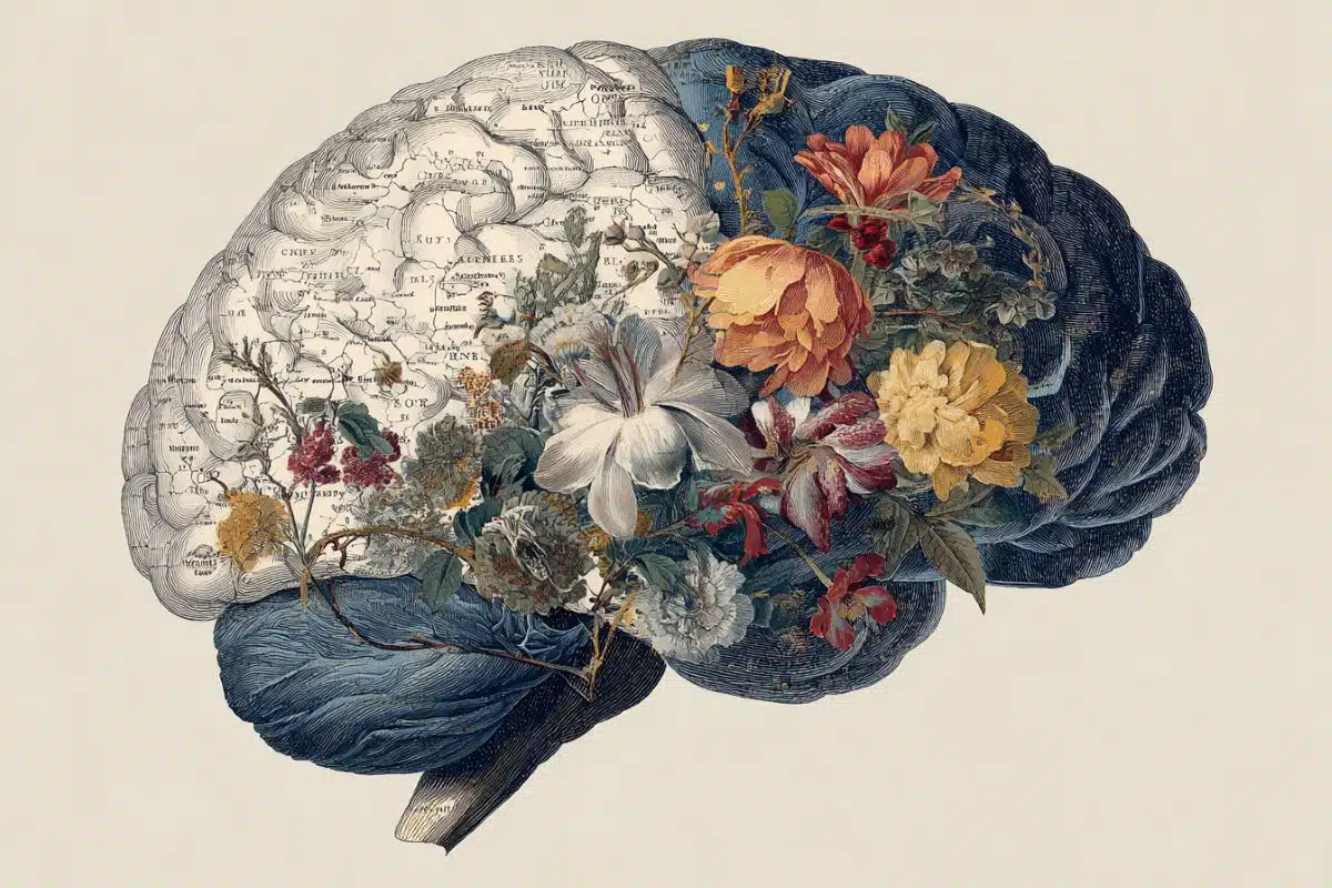
The process we call learning is in fact a well-orchestrated symphony of thousands of molecular reactions, but the exact interplay between these reactions remains largely unknown. Now, researchers at the Okinawa Institute of Science and Technology Graduate University (OIST) have modelled the molecular basis of learning in the cerebellum, a part of the brain that receives sensory input and coordinates voluntary movements.
“As far as we know, this is the most complex model of such a system that exists,” said Erik De Schutter, head of OIST’s Computational Neuroscience Unit and senior author on the recent paper, published in Cell Reports. Previous models focused on the signals that arrive at the receiving end of a neuron, he said, “whereas now we’re looking at the ongoing communication between the two ends.”
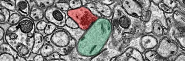
Learning is thought to be a balance between two processes that act as a kind of molecular dial: long-term potentiation (LTP), in which the connection between two neurons is strengthened, and long-term depression (LTD), in which the connection between two neurons is weakened. Both these processes take place at the synapse–the junction between two neurons. Andrew Gallimore, first author on the paper and a postdoctoral researcher at OIST, modeled how they work in two types of cells: parallel fibers and Purkinje cells, which play a key role in motor learning.
Using a computer program to create a model of this complex system, Gallimore combined several hundred equations taken from experiments in which such neurons were activated. The model was put to the test when colleagues in Korea took recordings from neurons in the cerebellum of mice. The OIST researchers then incorporated these recordings into the model.
Their findings show that the molecular networks on both sides of a synapse are important for controlling learning: communication must occur in both directions across the synapse to control whether LTD or LTP is generated during neural activity.
The model also showed that the molecular dial balancing LTP and LTD has an automatic off-switch that, when triggered, allows the system to return to its resting state. Although previous research hinted at the presence of this off-switch, this is the first time that the mechanism behind it–a complex network of proteins and receptors–has been demonstrated. Such a large, comprehensive model allows scientists to examine how complex signaling systems work together, something that is often absent in experimental literature, De Schutter said.
The researchers’ work allows scientists to more accurately predict the behavior of the chaotic, complex system of molecules that controls learning. It also hints at what might be happening at the molecular level when these switches break–which might occur when the brain is injured or during neurodegenerative diseases that affect learning.
“The whole function of a brain is based on the strengths of these synaptic connections,” said Gallimore. “The better we understand these processes, the greater potential there is to intervene to mitigate severe problems.”
Funding: Okinawa Institute of Science and Technology Graduate University, Korea Institute of Science and Technology Institutional Program funded this study.
Source: Kaoru Natori – OIST
Publisher: Organized by NeuroscienceNews.com.
Image Source: NeuroscienceNews.com image is credited to OIST Computational Neuroscience Unit.
Original Research: Open access research in Cell Reports.
doi:10.1016/j.celrep.2017.12.084
[cbtabs][cbtab title=”MLA”]OIST “Illuminating Mechanism at Play in Learning.” NeuroscienceNews. NeuroscienceNews, 15 March 2018.
<https://neurosciencenews.com/learning-mechanism-8645/>.[/cbtab][cbtab title=”APA”]OIST (2018, March 15). Illuminating Mechanism at Play in Learning. NeuroscienceNews. Retrieved March 15, 2018 from https://neurosciencenews.com/learning-mechanism-8645/[/cbtab][cbtab title=”Chicago”]OIST “Illuminating Mechanism at Play in Learning.” https://neurosciencenews.com/learning-mechanism-8645/ (accessed March 15, 2018).[/cbtab][/cbtabs]
Abstract
Switching On Depression and Potentiation in the Cerebellum
Highlights
•Unified bidirectional model of cerebellar parallel fiber-Purkinje cell LTD and LTP
•An ultrasensitive on/off switch mechanism demarcates the early phase of LTD
•A trans-synaptic feedback loop regulates nitric oxide production during LTD and LTP
•LTP’s frequency dependence determined by stimulus-dependent nitric oxide secretion
Summary
Long-term depression (LTD) and long-term potentiation (LTP) in the cerebellum are important for motor learning. However, the signaling mechanisms controlling whether LTD or LTP is induced in response to synaptic stimulation remain obscure. Using a unified model of LTD and LTP at the cerebellar parallel fiber-Purkinje cell (PF-PC) synapse, we delineate the coordinated pre- and postsynaptic signaling that determines the direction of plasticity. We show that LTP is the default response to PF stimulation above a well-defined frequency threshold. However, if the calcium signal surpasses the threshold for CaMKII activation, then an ultrasensitive “on switch” activates an extracellular signal-regulated kinase (ERK)-based positive feedback loop that triggers LTD instead. This postsynaptic feedback loop is sustained by another, trans-synaptic, feedback loop that maintains nitric oxide production throughout LTD induction. When full depression is achieved, an automatic “off switch” inactivates the feedback loops, returning the network to its basal state and demarcating the end of the early phase of LTD.