

May be possible to prune synapses with drug after diagnosis.
Children and adolescents with autism have a surplus of synapses in the brain, and this excess is due to a slowdown in a normal brain “pruning” process during development, according to a study by neuroscientists at Columbia University Medical Center (CUMC). Because synapses are the points where neurons connect and communicate with each other, the excessive synapses may have profound effects on how the brain functions. The study was published in the August 21 online issue of the journal Neuron.
A drug that restores normal synaptic pruning can improve autistic-like behaviors in mice, the researchers found, even when the drug is given after the behaviors have appeared.
“This is an important finding that could lead to a novel and much-needed therapeutic strategy for autism,” said Jeffrey Lieberman, MD, Lawrence C. Kolb Professor and Chair of Psychiatry at CUMC and director of New York State Psychiatric Institute, who was not involved in the study.
Although the drug, rapamycin, has side effects that may preclude its use in people with autism, “the fact that we can see changes in behavior suggests that autism may still be treatable after a child is diagnosed, if we can find a better drug,” said the study’s senior investigator, David Sulzer, PhD, professor of neurobiology in the Departments of Psychiatry, Neurology, and Pharmacology at CUMC.
During normal brain development, a burst of synapse formation occurs in infancy, particularly in the cortex, a region involved in autistic behaviors; pruning eliminates about half of these cortical synapses by late adolescence. Synapses are known to be affected by many genes linked to autism, and some researchers have hypothesized that people with autism may have more synapses.
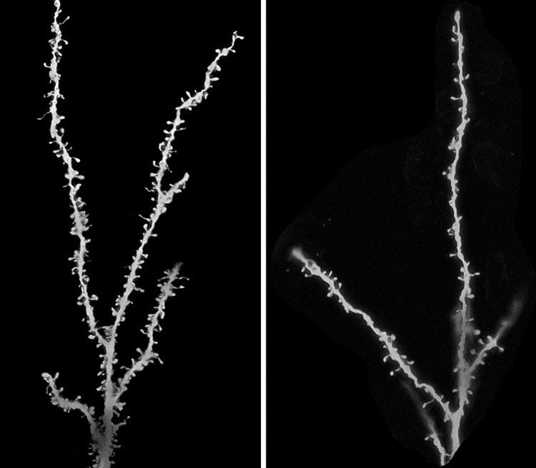
To test this hypothesis, co-author Guomei Tang, PhD, assistant professor of neurology at CUMC, examined brains from children with autism who had died from other causes. Thirteen brains came from children ages two to 9, and thirteen brains came from children ages 13 to 20. Twenty-two brains from children without autism were also examined for comparison.
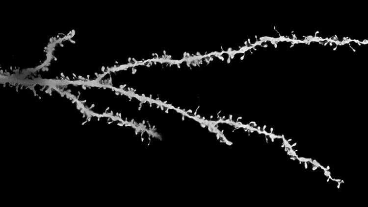
Dr. Tang measured synapse density in a small section of tissue in each brain by counting the number of tiny spines that branch from these cortical neurons; each spine connects with another neuron via a synapse.
By late childhood, she found, spine density had dropped by about half in the control brains, but by only 16 percent in the brains from autism patients.
“It’s the first time that anyone has looked for, and seen, a lack of pruning during development of children with autism,” Dr. Sulzer said, “although lower numbers of synapses in some brain areas have been detected in brains from older patients and in mice with autistic-like behaviors.”
Clues to what caused the pruning defect were also found in the patients’ brains; the autistic children’s brain cells were filled with old and damaged parts and were very deficient in a degradation pathway known as “autophagy.” Cells use autophagy (a term from the Greek for self-eating) to degrade their own components.
Using mouse models of autism, the researchers traced the pruning defect to a protein called mTOR. When mTOR is overactive, they found, brain cells lose much of their “self-eating” ability. And without this ability, the brains of the mice were pruned poorly and contained excess synapses. “While people usually think of learning as requiring formation of new synapses, “Dr. Sulzer says, “the removal of inappropriate synapses may be just as important.”
The researchers could restore normal autophagy and synaptic pruning—and reverse autistic-like behaviors in the mice—by administering rapamycin, a drug that inhibits mTOR. The drug was effective even when administered to the mice after they developed the behaviors, suggesting that such an approach may be used to treat patients even after the disorder has been diagnosed.
Because large amounts of overactive mTOR were also found in almost all of the brains of the autism patients, the same processes may occur in children with autism.
“What’s remarkable about the findings,” said Dr. Sulzer, “is that hundreds of genes have been linked to autism, but almost all of our human subjects had overactive mTOR and decreased autophagy, and all appear to have a lack of normal synaptic pruning. This says that many, perhaps the majority, of genes may converge onto this mTOR/autophagy pathway, the same way that many tributaries all lead into the Mississippi River. Overactive mTOR and reduced autophagy, by blocking normal synaptic pruning that may underlie learning appropriate behavior, may be a unifying feature of autism.”
Alan Packer, PhD, senior scientist at the Simons Foundation, which funded the research, said the study is an important step forward in understanding what’s happening in the brains of people with autism.

“The current view is that autism is heterogeneous, with potentially hundreds of genes that can contribute. That’s a very wide spectrum, so the goal now is to understand how those hundreds of genes cluster together into a smaller number of pathways; that will give us better clues to potential treatments,” he said.
“The mTOR pathway certainly looks like one of these pathways. It is possible that screening for mTOR and autophagic activity will provide a means to diagnose some features of autism, and normalizing these pathways might help to treat synaptic dysfunction and treat the disease.”
The paper is titled, “Loss of mTOR-dependent macroautophagy causes autistic-like synaptic pruning deficits.” Other authors are: Kathryn Gudsnuk, Sheng-Han Kuo, Marisa L. Cotrina, Gorazd Rosoklija, AlexanderSosunov, Mark S. Sonders, Ellen Kanter, Candace Castagna, Ai Yamamoto, OttavioArancio, Bradley S. Peterson, Frances Champagne, Andrew J. Dwork, and James Goldman from CUMC; and Zhenyu Yue (Icahn School of Medicine at Mount Sinai). Marisa Cotrina is now at the University of Rochester.
The authors declare no competing financial interests.
This study was supported by the Simons Foundation, with additional funding from the U.S. Department of Defense (TS110056), the Parkinson’s Disease Foundation, JPB Foundation, National Institutes of Health (K01MH096956, R01MH64168, DP2OD001674, R01NS049442), and the American Heart Association. Harvard Brain Bank and Maryland NICHD Brain & Tissue Bank provided brain tissue.
Contact: Karin Eskenazi – Columbia University Medical Center
Source: Columbia University Medical Center press release
Image Source: The images are credited to Guomei Tang and Mark S. Sonders, and are adapted from the Columbia University Medical Center press release
Original Research: Full open access research for “Loss of mTOR-Dependent Macroautophagy Causes Autistic-like Synaptic Pruning Deficits” by Guomei Tang, Kathryn Gudsnuk, Sheng-Han Kuo, Marisa L. Cotrina, Gorazd Rosoklija, Alexander Sosunov, Mark S. Sonders, Ellen Kanter, Candace Castagna, Ai Yamamoto, Zhenyu Yue, Ottavio Arancio, Bradley S. Peterson, Frances Champagne, Andrew J. Dwork, James Goldman, and David Sulzer in Neuron. Published online August 21 2014 doi:10.1016/j.neuron.2014.07.040


