

Summary: Researchers have succeeded in making superficial white matter visible in a living human brain.
Source: Max Planck Institute
Traditionally, neuroscience regards the brain as being made up of two basic tissue types. Billions of neurons make up the grey matter, forming a thin layer on the brain’s surface. These neuronal cells are interlinked in a mindboggling network by hundreds of millions of white matter connections, running in bundles, deeper in the brain.
Until very recently, not much was known about the interface between the white and grey matter – the so-called superficial white matter – because methods were lacking to study it in living human brains. Yet, previous investigations had suggested the region to be implicated in devastating conditions such as Alzheimer’s disease and autism.
Now a multidisciplinary team led by Nikolaus Weiskopf from the Max Planck Institute for Human Cognitive and Brain Sciences has succeeded in making the superficial white matter visible in the living human brain.
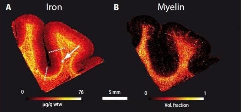
“We demonstrated that the superficial white matter contains a lot of iron. It is known that iron is necessary for the process of myelination,” explains Evgeniya Kirilina, first author of the study published in Science Advances.
Myelin is what makes the white matter white. It’s the fatty coating of nerve cell axons that speeds up transmission of information through the brain. The myelination process can occur throughout the lifespan but is predominant during development. In fact, the largest concentration of iron the researchers found was in the superficial white matter in regions of the frontal cortex, which happens to be the slowest developing structure in the human brain. Incredibly, the human frontal cortex is not fully myelinated until the forth decade of life.
The key to the new method is MRI (Magnetic Resonance Imaging) but at very high field strength. While typical clinical MRI scanners work at 1.5 or 3 Tesla, in terms of the strength of the magnetic field, the Max Planck Institute for Human Cognitive and Brain Sciences houses a powerful 7 Tesla scanner.
This, in combination with advanced biophysical model, allowed the team to create very high resolution maps of the white-grey matter border across the entire living brain. The accuracy of their submillimetre maps was assessed against classic and advanced histological methods involving physical dissection and analysis of post mortem brains.
The new method promises many further insights into the organisation of the interface between white and grey matter. Evgeniya Kirilina adds, “We hope the method can be used to increase our understanding of brain development as well as pathological conditions involving the superficial white matter.”
About this neuroscience research news
Source: Max Planck Institute
Contact: Bettina Hennebach – Max Planck Institute
Image: Image is credited to Max Planck Institute
Original Research: Open access.
“Superficial white matter imaging: Contrast mechanisms and whole-brain in vivo mapping ” by Evgeniya Kirilina, Saskia Helbling, Markus Morawski, Kerrin Pine, Katja Reimann, Steffen Jankuhn, Juliane Dinse, Andreas Deistung, Jürgen R. Reichenbach, Robert Trampel, Stefan Geyer, Larissa Müller, Norbert Jakubowski, Thomas Arendt, Pierre-Louis Bazin, Nikolaus Weiskopf . Science Advances
Abstract
Superficial white matter imaging: Contrast mechanisms and whole-brain in vivo mapping
Superficial white matter (SWM) contains the most cortico-cortical white matter connections in the human brain encompassing the short U-shaped association fibers. Despite its importance for brain connectivity, very little is known about SWM in humans, mainly due to the lack of noninvasive imaging methods. Here, we lay the groundwork for systematic in vivo SWM mapping using ultrahigh resolution 7 T magnetic resonance imaging. Using biophysical modeling informed by quantitative ion beam microscopy on postmortem brain tissue, we demonstrate that MR contrast in SWM is driven by iron and can be linked to the microscopic iron distribution. Higher SWM iron concentrations were observed in U-fiber–rich frontal, temporal, and parietal areas, potentially reflecting high fiber density or late myelination in these areas. Our SWM mapping approach provides the foundation for systematic studies of interindividual differences, plasticity, and pathologies of this crucial structure for cortico-cortical connectivity in humans.


