

Researchers have discovered a thick band of microtubules in certain neurons in the retina that they believe acts as a transport road for mitochondria that help provide energy required for visual processing. The findings appear in the July issue of The Journal of General Physiology.
The retina is a layer of tissue in the back of the eye that converts light into nerve impulses. The retina contains small, specialized neurons called bipolar cells that transmit information from light-sensitive photoreceptor cells to ganglion neurons, which send information to the brain for interpretation as images.
Bipolar cells are continuously active, a characteristic few other neurons share. They require a constant supply of energy to mediate the sustained release of the contents of an enormous number of synaptic vesicles, which store the transmitters that convey information between neurons. An intriguing new study of their subcellular structure could help explain how bipolar synaptic terminals meet such excessive energy demands.
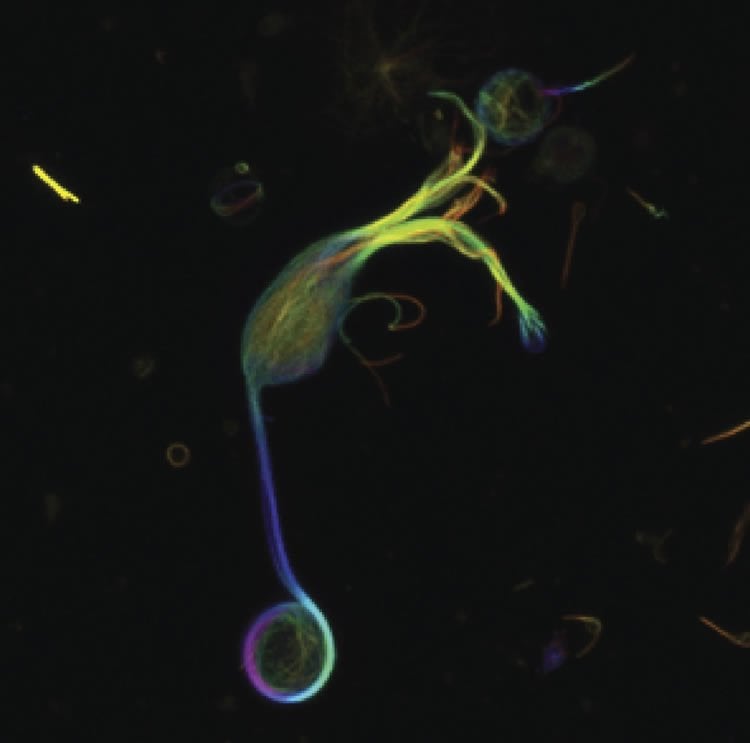
Using cutting-edge 3D microscopy, researchers from the National Heart, Lung, and Blood Institute and Yale University examined the subcellular architecture of presynaptic terminals in retinal bipolar cells of live goldfish. Goldfish retinal bipolar cells have giant presynaptic terminals that make them especially amenable for investigation. Unexpectedly, the team discovered a thick band of microtubules, a component of the cell’s cytoskeleton, that extended from the axon of the neuron into the synaptic terminal and then looped around the interior periphery of the terminal.
The microtubule band appeared to associate with mitochondria–organelles known for providing energy to cells–in the synaptic terminal. When the researchers administered drugs to inhibit the movement of certain “motor” proteins that transport mitochondria and other cargo within the cell by traveling along microtubules, the mitochondria accumulated in the axon of the neuron and never made it to the synaptic terminal.
The findings suggest that these previously unknown microtubule structures provide a “roadway” for the transport of mitochondria crucial to maintain energy supplies into the synaptic terminals of these highly active neurons associated with vision.
Funding: The funding was provided by the NIH/National Heart Lung and Blood Institute, National Institutes of Health.
Source: Rita Sullivan King – Rockefeller University Press
Image Credit: Image is credited to Graffe et al.
Original Research: Abstract for “A marginal band of microtubules transports and organizes mitochondria in retinal bipolar synaptic terminals” by Malkolm Graffe, David Zenisek, and Justin W. Taraska in Journal of General Physiology. Published online June 29 2015 doi:10.1085/jgp.201511396
Abstract
A marginal band of microtubules transports and organizes mitochondria in retinal bipolar synaptic terminals
A set of bipolar cells in the retina of goldfish contains giant synaptic terminals that can be over 10 µm in diameter. Hundreds of thousands of synaptic vesicles fill these terminals and engage in continuous rounds of exocytosis. How the cytoskeleton and other organelles in these neurons are organized to control synaptic activity is unknown. Here, we used 3-D fluorescence and 3-D electron microscopy to visualize the complex subcellular architecture of these terminals. We discovered a thick band of microtubules that emerged from the axon to loop around the terminal periphery throughout the presynaptic space. This previously unknown microtubule structure associated with a substantial population of mitochondria in the synaptic terminal. Drugs that inhibit microtubule-based kinesin motors led to accumulation of mitochondria in the axon. We conclude that this prominent microtubule band is crucial to the transport and localization of mitochondria into the presynaptic space to provide the sustained energy necessary for continuous transmitter release in these giant synaptic terminals.
“When you smile, the world smiles at you: ERP evidence for self-expression effects on face processing” by Alejandra Sel, Beatriz Calvo-Merino, Simone Tuettenberg and Bettina Forster in Social Cognitive and Affective Neuroscience. Published online June 2015 doi:10.1093/scan/nsv009


