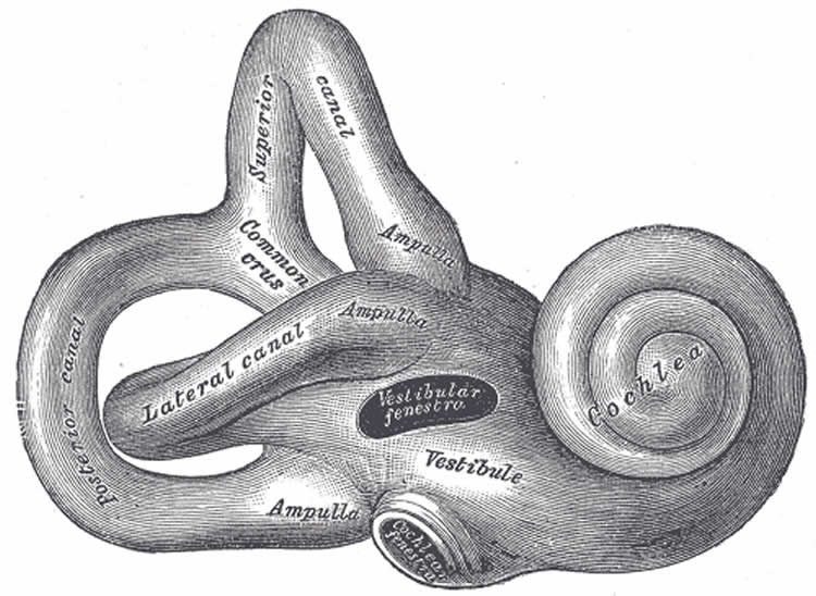
Neurologists at LMU have studied the role of the vestibular system, which controls balance, in optimizing how we direct our gaze. The results could lead to more effective rehabilitation of patients with vestibular or cerebellar dysfunction.
When we shift the direction of our gaze, head and eye movements are normally highly coordinated with each other. Indeed, from the many possible combinations of speed and duration for such movements, the brain chooses the one that minimizes the error in reaching the intended line of sight. Dr. Nadine Lehnen, who heads a research group based at LMU’s Center for Vertigo and Balance Disorders, in collaboration with her colleague Dr. Murat Saglam and Professor Stefan Glasauer of the Center for Sensorimotor Diseases at LMU, have now published a paper in the latest issue of the journal of Brain which investigates the significance of the vestibular system for this optimization of motor coordination. The vestibular system in the brain is mainly responsible for the maintenance of balance and posture. The new work focused on subjects suffering from bilateral defects in the vestibular system (a complete vestibulopathy) or lesions in the cerebellum, which is functionally linked to it.
The authors of the new study had previously developed a mathematical model that enabled them to predict the horizontal movements of the head and eyes in response to the presentation of an off-center stimulus. “When subjected to repeated trials, healthy subjects are able to select the combination of eye and head movements that minimizes gaze shift variability,” says Glasauer. They unconsciously choose the set of movements associated with the least error in the endpoint. Moreover, they can do this even when wearing a helmet with weights attached, which alters the moment of inertia of the head.

Learning to find the endpoint
However, patients who show defects in the vestibular system or the cerebellum have greater difficulty in controlling the direction of gaze in response to changes in their environment. “It turns out that information relayed from the balance organs to the vestibular system is essential for the optimization of gaze shifts,” says Nadine Lehnen. Patients with complete bilateral vestibular loss are therefore unable to perform such shifts in the most efficient way. “In striking contrast, patients with cerebellar damage can, to a certain extent, learn to optimize certain parameters of head and eye movements, by adjusting the velocity of head movement, for instance,” says Glasauer.
“These results provide the first evidence that the vestibular system is critical for optimizing voluntary movements“, says Dr. Kathleen E. Cullen from McGill University in Montreal in a scientific commentary to the study appearing in the print issue of Brain. The new findings are of relevance for the rehabilitation of patients who have suffered damage to the cerebellum and patients with incomplete vestibulopathies.
“We assume that gaze shift control in these patients can be enhanced by a rehabilitation training based on active head movements,” says Nadine Lehnen. Head movements provide the vestibular feedback which generates the sensorimotor error messages that underlie the ability to learn how to optimize the coordination of eye and head movements. Instead of trying to hold their heads steady, these patients should be encouraged to actively move their heads, when they shift their gaze.
The question if patients with partial vestibulopathy can optimize gaze shift behavior by engaging in active head movements is now under investigation. This work forms part of a rehabilitation study which is being carried out at the Center for Vertigo and Balance Disorders at Munich University Hospitals, and is financed by the Federal Ministry for Education and Research.
Contact: Luise Dirscherl – Ludwig Maximilians University, Munich
Source: Ludwig Maximilians University, Munich press release
Image Source: The image is credited to Gray’s Anatomy and is in the public domain.
Original Research: Abstract for “Vestibular and cerebellar contribution to gaze optimality” by Murat Sağlam, Stefan Glasauer and Nadine Lehnen in Brain. Published online February 17 2014 doi:10.1093/brain/awu006


