

New optical technique, used in mice, offers detailed look at how dopamine works in the brain.
Columbia University scientists have developed a new optical technique to study how information is transmitted in the brains of mice. Using this method, they found that only a small portion of synapses — the connections between cells that control brain activity–may be active at any given time.
The study was published in the latest issue of Nature Neuroscience.
“Understanding how we accomplish complex tasks, such as learning and memory, requires us to look at how our brains transmit key signals — called neurotransmitters — across synapses from one neuron to another,” said David Sulzer, PhD, professor of neurobiology in Psychiatry, Neurology, and Pharmacology at Columbia University Medical Center (CUMC). “Older techniques only revealed what was going on in large groups of synapses. We needed a way to observe the neurotransmitter activity of individual synapses, to help us better understand their intricate behavior.”
To obtain a detailed view of synaptic activity, Sulzer’s team collaborated with the laboratory of Dalibor Sames, PhD, associate professor of chemistry at Columbia, to develop a novel compound called fluorescent false neurotransmitter 200 (FFN200). When added to brain tissue or nerve cells from mice, FFN200 mimics the brain’s natural neurotransmitters and allows researchers to spy on chemical messaging in action.
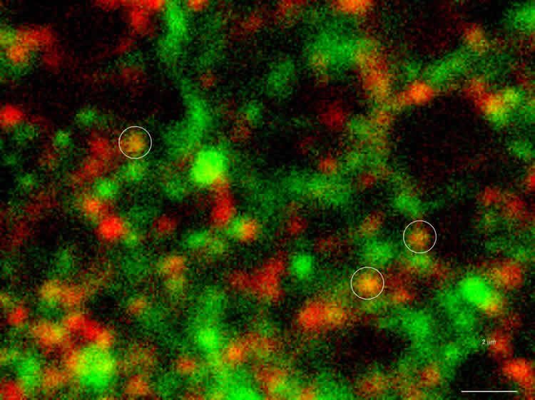
Using a fluorescence microscope, the researchers were able to view the release and reuptake of dopamine — a neurotransmitter involved in motor learning, habit formation, and reward-seeking behavior — in individual synapses. When all the neurons were electrically stimulated in a sample of brain tissue, the researchers expected all the synapses to release dopamine. Instead, they found that less than 20 percent of dopaminergic synapses were active following a pulse of electricity.
“Why are there these large reservoirs of synapses that are silent?” said Dr. Sames, a co-author of the paper. “Perhaps these silent terminals hint at a mechanism of information coding in the brain that’s yet to be revealed.”
The study’s authors plan to pursue this hypothesis in future experiments, as well as examine how other neurotransmitters behave.
Columbia University scientists have developed a new optical technique to study how information is transmitted in the brains of mice. Using this method, they found that only a small portion of synapses—the connections between cells that control brain activity—may be active at any given time.
“This particular study didn’t explain what’s causing most of the synapses to remain silent,” said Dr. Sulzer. “If we can work this out, we may learn a lot more about how alterations in dopamine levels are involved in brain disorders such as Parkinson’s disease, addiction, and schizophrenia.”
The study is titled, “Fluorescent false neurotransmitter reveals functionally silent dopamine vesicle clusters in the striatum.” The other contributors are: Daniela B. Pereira, Yvonne Schmitz, Josef Meszaros, Paolomi Merchant, Gang Hu, Shu Li, Adam Henke, Jose E. Lizardi-Ortiz, Richard J. Karpowicz, Jr., Travis J. Morgenstern, Mark S. Sonders, Ellen Kanter, Pamela C. Rodriguez, and Eugene V. Mosharov,.
Funding: The study was supported by grants from the G. Harold & Leila Y. Mathers Charitable Foundation, the National Institute of Mental Health (grants R01MH086545 and R01MH108186), the National Institute of Neurological Disorders and Stroke (grant R01NS075222), the National Science Foundation, the JPB, McKnight and Michael J. Fox Foundation, and the Udall Center of Excellence for Parkinson’s Disease Research.
The Parkinson’s Disease Foundation also supported this work.
The authors declare competing financial interests: details are available in the online version of the paper. D. Sulzer and D. Sames were listed as inventors on a patent (8,337,941) covering FFN200 and a patent application (13/575,535) covering FFN102, com- pounds employed in this study.
Source: Lucky Tran, PhD – Columbia University Medical Center
Image Credit: Image is credited to Sulzer Lab/Columbia University Medical Center.
Video Source: The video is credited to Columbia Medical Center.
Original Research: Abstract for “Fluorescent false neurotransmitter reveals functionally silent dopamine vesicle clusters in the striatum” by Daniela B Pereira, Yvonne Schmitz, József Mészáros, Paolomi Merchant, Gang Hu, Shu Li, Adam Henke, José E Lizardi-Ortiz, Richard J Karpowicz Jr, Travis J Morgenstern, Mark S Sonders, Ellen Kanter, Pamela C Rodriguez, Eugene V Mosharov, Dalibor Sames and David Sulzer in Nature Neuroscience. Published online February 22 2016 doi:10.1038/nn.4252
Abstract
Fluorescent false neurotransmitter reveals functionally silent dopamine vesicle clusters in the striatum
Neurotransmission at dopaminergic synapses has been studied with techniques that provide high temporal resolution, but cannot resolve individual synapses. To elucidate the spatial dynamics and heterogeneity of individual dopamine boutons, we developed fluorescent false neurotransmitter 200 (FFN200), a vesicular monoamine transporter 2 (VMAT2) substrate that selectively traces monoamine exocytosis in both neuronal cell culture and brain tissue. By monitoring electrically evoked Ca2+ transients with GCaMP3 and FFN200 release simultaneously, we found that only a small fraction of dopamine boutons that exhibited Ca2+ influx engaged in exocytosis, a result confirmed with activity-dependent loading of the endocytic probe FM1-43. Thus, only a low fraction of striatal dopamine axonal sites with uptake-competent VMAT2 vesicles are capable of transmitter release. This is consistent with the presence of functionally ‘silent’ dopamine vesicle clusters and represents, to the best of our knowledge, the first report suggestive of presynaptically silent neuromodulatory synapses.
“Fluorescent false neurotransmitter reveals functionally silent dopamine vesicle clusters in the striatum” by Daniela B Pereira, Yvonne Schmitz, József Mészáros, Paolomi Merchant, Gang Hu, Shu Li, Adam Henke, José E Lizardi-Ortiz, Richard J Karpowicz Jr, Travis J Morgenstern, Mark S Sonders, Ellen Kanter, Pamela C Rodriguez, Eugene V Mosharov, Dalibor Sames and David Sulzer in Nature Neuroscience. Published online February 22 2016 doi:10.1038/nn.4252


