

Carnegie Mellon University neuroscientists have identified a new pathway by which several brain areas communicate within the brain’s striatum.
Published in the Journal of Neuroscience, the findings illustrate structural and functional connections that allow the brain to use reinforcement learning to make spatial decisions, such as the dorsolateral prefrontal (DLPFC), orbitofrontal cortex (OFC) and posterior parietal cortex (PPC). Communication between these regions is important for abilities like how a baseball player is able to estimate where to swing his bat or how a person finds a car in a large parking lot filled with similar cars.
Knowing how these specific pathways work together provides crucial insight into how learning occurs. It also could lead to improved treatments for Parkinson’s disease.
“By understanding precisely how these systems communicate together, we can come up with a better understanding for how these systems operate in the healthy brain, but also start to understand how in Parkinson’s disease different types of systems ‘cascade,’ or start with one symptom like motor dysfunction and move to another like memory or decision-making problems,” said Timothy Verstynen, assistant professor of psychology and a faculty member in the Center for the Neural Basis of Cognition (CNBC) in CMU’s Dietrich College of Humanities and Social Sciences.
The hope is that more knowledge of how the connectivity is related to behavior will help scientists develop therapeutic interventions that focus on strengthening potentially weakened or damaged pathways.
For the study, Verstynen and Kevin Jarbo, a Ph.D. student in psychology, used diffusion spectrum imaging and fiber technology to analyze brain images collected from 60 healthy adults. The advanced imaging techniques allowed Verstynen and Jarbo to visualize the white matter pathways from the DLPFC, OFC and PPC.
They found that the pathways from all three areas projected to similar areas within a forebrain region called striatum, a part of the basal ganglia pathways that are most commonly associated with Parkinson’s disease. The patterns were consistent across all participants.
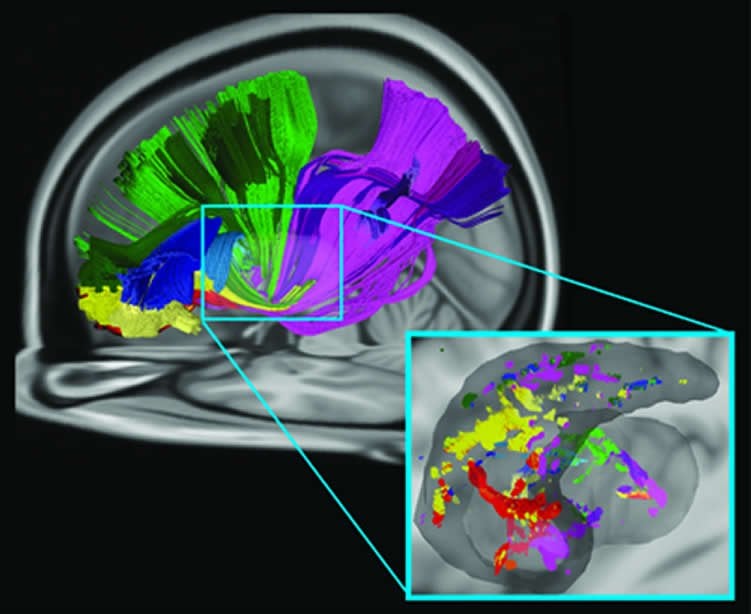
The researchers followed the structural connectivity analysis with a functional connectivity analysis by using resting state fMRI images. The results showed that the convergence zones were not only structurally connected but functionally connected as well. More importantly, the areas at the surface of the brain in all three cortical areas showed a high overlap of structure and functional connectivity.
“Our findings suggest that there may be a structural and functional network in the brain that allows us to integrate information about where we are focusing our attention in our visuospatial environment with reward and punishment signals associated with our past action choices in order to learn how to update, and hopefully improve, our future action decisions,” Jarbo said.
An additional implication for this study is a deeper understanding of how reinforcement learning occurs.
“A lot of models of the reinforcement learning process assume that reward signals from the orbitofrontal cortex converge with information from other areas. These have been shown to be true for other regions of the prefrontal cortex. We are the first to show that spatial attention information from the parietal cortex may also contribute to this process,” Verstynen said.
The Pennsylvania Department of Health, the Department of Defense and the National Institutes of Health funded this research.
Contact: Shilo Rea – Carnegie Mellon University
Source: Carnegie Mellon University press release
Image Source: The image is adapted from the Carnegie Mellon University press release
Original Research: Abstract for “Converging Structural and Functional Connectivity of Orbitofrontal, Dorsolateral Prefrontal, and Posterior Parietal Cortex in the Human Striatum” by Kevin Jarbo and Timothy D. Verstynen in Journal of Neuroscience. Published online March 5 2015 doi:10.1523/JNEUROSCI.2636-14.2015


