

Summary: According to researchers, the adult brain may be sensitive to social and economic factors. Researchers report in middle age, better socioeconomic status is associated with more efficient brain network organization and thicker gray matter.
Source: UT Dallas.
Research has shown that a developing child’s brain structure and function can be adversely affected when the child is raised in an environment lacking adequate education, nutrition and access to health care.
While the impact of such an environment on children is relatively well understood, a new study from The University of Texas at Dallas examines an effect that is not so clear — the relationship of socioeconomic status (SES) to brain function and anatomy in adults.
The study, led by researchers at the Center for Vital Longevity at UT Dallas, found that the adult brain may actually be sensitive to social and economic factors.
“We know that socioeconomic status influences the structure of the brain in childhood and older age, but there’s been a gap in the research. We wanted to see if there were relationships between SES and the brain across a wider range of adulthood,” said Dr. Gagan Wig, assistant professor in the School of Behavioral and Brain Sciences at UT Dallas and corresponding author of the study published online in the Proceedings of the National Academy of Sciences.
The study included 304 individuals ranging between 20 and 89 years of age. Each individual’s SES was approximated using standard methods that combine education and a measure of occupational prestige. The SES measure was also correlated with individual income and reports of subjective SES standing.
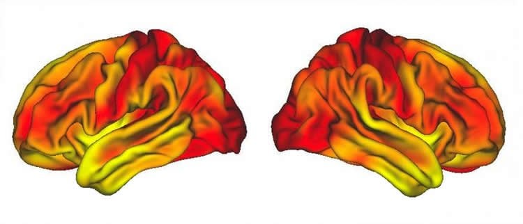
To measure brain function, the researchers used functional magnetic resonance imaging to collect a type of brain scan that shows how an individual’s functional brain networks are organized while the participant lies in the machine in a state of resting wakefulness. In addition, the researchers used anatomical brain scans to measure the thickness of cortical grey matter in each individual’s brain.
In middle-age adults, ages 35 to 64 years, a higher socioeconomic status was associated with more efficiently organized brain networks and thicker cortical grey matter. Those who ranked lower in SES tended to have less well-organized functional brain networks and a thinner cortex. A thinner cortex can contribute to cognitive impairment later in life, such as memory loss and dementia.
“We know less about the impact of brain network organization on later life outcomes, but these results suggest that it is worth further study,” Wig said.
The SES-brain relationships persisted after controlling for demographics, measures of both physical and mental health, and cognitive ability. In addition, the relationship between an individual’s present adult SES and their brain-network organization was independent of his or her childhood SES, providing evidence that the SES relationships are not simply due to differences that have been previously established during the earliest years of life.
The relationship between SES and the brain measures was diminished in the elderly. The scientists suggest that this may be due to the fact that older age can be associated with greater brain changes that obscure any SES relationships.
The scientists caution that more research is needed to gain a better understanding of potential relationships between socioeconomic factors and brain health.
“These data provide a snapshot in time for each participant,” said the study’s lead author Dr. Micaela Chan, a postdoctoral researcher working with Wig at the Center for Vital Longevity. “Following individuals through their lifespan would provide more information about brain changes and their relationship to life events and status.”
“What we have found in middle-aged adults is a correlation between socioeconomic status and brain function and anatomy,” Wig said. “What makes these results more striking is that the individuals we studied were predominantly above the poverty line. This provides evidence that SES-brain relationships are not limited to individuals at the extreme ends of SES, but are present across a broader SES range. However, because differences in SES can be associated with differences in many factors, including those related to diet and health behaviors, access to health care, and levels of stress, it’s not yet clear which of these, if any, is the source of the observed relationships.
“The bottom line is, socioeconomic status might matter for brain health, even in middle age, and we will need to investigate the relationship further,” he said.
Participants were recruited from the Dallas-Fort Worth community through the Dallas Lifespan Brain Study, a brain-aging study started and led by Dr. Denise Park, also at the Center for Vital Longevity and a contributing author of the study. Other authors include Phillip Agres, a doctoral student in the School of Behavioral and Brain Sciences, Neil Savalia, a research assistant in Wig’s laboratory, and Dr. Jinkyung Na, associate professor at Sogang University in Seoul.
Funding: The work was supported by the National Institute on Aging – part of the National Institutes of Health – and the James S. McDonnell Foundation.
Source: Amanda Siegfried – UT Dallas
Publisher: Organized by NeuroscienceNews.com.
Image Source: NeuroscienceNews.com image is credited to Dr. Gagan Wig, Center for Vital Longevity, University of Texas at Dallas.
Original Research: Open access research for “Socioeconomic status moderates age-related differences in the brain’s functional network organization and anatomy across the adult lifespan” by Micaela Y. Chan, Jinkyung Na, Phillip F. Agres, Neil K. Savalia, Denise C. Park, and Gagan S. Wig in PNAS. Published May 4 2018.
doi:10.1073/pnas.1714021115
[cbtabs][cbtab title=”MLA”]UT Dallas “Relationship Between Socioeconomic Status and the Adult Brain.” NeuroscienceNews. NeuroscienceNews, 15 May 2018.
<https://neurosciencenews.com/socioeconomic-adult-brain-9057/>.[/cbtab][cbtab title=”APA”]UT Dallas (2018, May 15). Relationship Between Socioeconomic Status and the Adult Brain. NeuroscienceNews. Retrieved May 15, 2018 from https://neurosciencenews.com/socioeconomic-adult-brain-9057/[/cbtab][cbtab title=”Chicago”]UT Dallas “Relationship Between Socioeconomic Status and the Adult Brain.” https://neurosciencenews.com/socioeconomic-adult-brain-9057/ (accessed May 15, 2018).[/cbtab][/cbtabs]
Abstract
Socioeconomic status moderates age-related differences in the brain’s functional network organization and anatomy across the adult lifespan
An individual’s environmental surroundings interact with the development and maturation of their brain. An important aspect of an individual’s environment is his or her socioeconomic status (SES), which estimates access to material resources and social prestige. Previous characterizations of the relation between SES and the brain have primarily focused on earlier or later epochs of the lifespan (i.e., childhood, older age). We broaden this work to examine the relationship between SES and the brain across a wide range of human adulthood (20–89 years), including individuals from the less studied middle-age range. SES, defined by education attainment and occupational socioeconomic characteristics, moderates previously reported age-related differences in the brain’s functional network organization and whole-brain cortical structure. Across middle age (35–64 years), lower SES is associated with reduced resting-state system segregation (a measure of effective functional network organization). A similar but less robust relationship exists between SES and age with respect to brain anatomy: Lower SES is associated with reduced cortical gray matter thickness in middle age. Conversely, younger and older adulthood do not exhibit consistent SES-related difference in the brain measures. The SES–brain relationships persist after controlling for measures of physical and mental health, cognitive ability, and participant demographics. Critically, an individual’s childhood SES cannot account for the relationship between their current SES and functional network organization. These findings provide evidence that SES relates to the brain’s functional network organization and anatomy across adult middle age, and that higher SES may be a protective factor against age-related brain decline.


