

Summary: A new study reports sensory stimulation during the first two weeks of life is critical for establishing brain networks that help map sensation.
Source: Weill Cornell Medicine.
Sensory stimulation during a critical period soon after birth is essential for establishing networks in the brain that “map” sensations and enable the development of normal behaviors, according to a new study by Weill Cornell Medicine investigators.
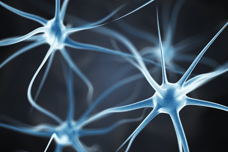
The paper, published in print July 11 and online June 19 in Neuron, is the first to observe the early functional development of specialized nerve cells, called interneurons, in mice during the first two weeks after birth. The findings suggest that there may be an early critical window for the development of interneurons in human babies for the establishment of healthy sensory responses in adulthood.
“How and when interneurons develop is important to understand because increasing evidence links abnormal interneuron activity to disorders such as autism and childhood epilepsy,” said Dr. Natalia De Marco Garcia, an assistant professor of neuroscience in the Feil Family Brain and Mind Research Institute at Weill Cornell Medicine. “If the neural circuits needed for responding to sensory inputs do not form correctly in early life, the deficits can lead to abnormal responses to sensory inputs and impaired social relationships in adults.”
Baby mice are unable to move their whiskers at birth. External stimulation through touch by the mother and their siblings sends messages from the whiskers to the brain. Baby mice gain the ability to move their whiskers voluntarily after interneurons develop and connect sensory and central neurons in a network to “map” the whiskers onto a specific area of the brain, joining sensory input and muscle movement.
For the study, Dr. De Marco Garcia and her team used puffs of air on the baby mice’s whiskers to simulate touch sensations that occur in the nest. Using specialized imaging technology, they identified the area of the brain responsible for receiving whisker-touch messages. They observed daily changes in the activity of single neurons as the mice matured and discovered that interneurons functionally developed as early as six days after birth.
Next, the researchers examined baby mice that were not able to fully experience touch sensations through the whiskers. As the mice grew and developed into adults, they still received sensory messages in the same brain areas as normal mice. However, they showed an impaired ability to distinguish textures by touch, indicating that they were not processing sensory messages correctly. “We demonstrated that perturbing the development of sensory networks during the critical window led to long-lasting behavioral deficits in mice,” Dr. De Marco Garcia said.
The findings shed light on how sensory-cognitive deficits may develop in disorders that arise during the development of the human nervous system, such as autism spectrum disorder (ASD). Many children with ASD have an impaired ability to process signals to the brain provided by the senses and respond abnormally to tactile, auditory and visual stimuli. Currently, the average age of diagnosis for ASD is around 4 years old with the disorders affecting an estimated 1 in about 59 children in the United States. Identifying the earliest clues of ASD is essential since early diagnosis and intervention can greatly improve a child’s development.
“Our findings pave the way for further research on how neurons develop in the critical window of early post-natal life in mouse models of autism,” Dr. De Marco Garcia said. “In the future, if we could observe impairments in interneuron activity in children at earlier stages of life, we might be able to intervene sooner and improve outcomes.”
Source: Weill Cornell Medicine
Publisher: Organized by NeuroscienceNews.com.
Image Source: NeuroscienceNews.com image is adapted from the Weill Cornell Medicine news release.
Original Research: Abstract for “Layer I Interneurons Sharpen Sensory Maps during Neonatal Development” by Alicia Che, Rachel Babi, jAndrew F. Iannone, Robert N. Fetcho, Monica Ferrer, Conor Liston, Gord Fishell, and Natalia V. De Marco García in Neuron. Published June 21 2018.
doi:10.1016/j.neuron.2018.06.002
[cbtabs][cbtab title=”MLA”]Weill Cornell Medicine”Early Sensory Stimulation is Critical for the Development of Sensory Networks.” NeuroscienceNews. NeuroscienceNews, 11 August 2018.
<https://neurosciencenews.com/sensory-stimulation-network-9690/>.[/cbtab][cbtab title=”APA”]Weill Cornell Medicine(2018, August 11). Early Sensory Stimulation is Critical for the Development of Sensory Networks. NeuroscienceNews. Retrieved August 11, 2018 from https://neurosciencenews.com/sensory-stimulation-network-9690/[/cbtab][cbtab title=”Chicago”]Weill Cornell Medicine”Early Sensory Stimulation is Critical for the Development of Sensory Networks.” https://neurosciencenews.com/sensory-stimulation-network-9690/ (accessed August 11, 2018).[/cbtab][/cbtabs]
Abstract
Layer I Interneurons Sharpen Sensory Maps during Neonatal Development
The neonatal mammal faces an array of sensory stimuli when diverse neuronal types have yet to form sensory maps. How these inputs interact with intrinsic neuronal activity to facilitate circuit assembly is not well understood. By using longitudinal calcium imaging in unanesthetized mouse pups, we show that layer I (LI) interneurons, delineated by co-expression of the 5HT3a serotonin receptor (5HT3aR) and reelin (Re), display spontaneous calcium transients with the highest degree of synchrony among cell types present in the superficial barrel cortex at postnatal day 6 (P6). 5HT3aR Re interneurons are activated by whisker stimulation during this period, and sensory deprivation induces decorrelation of their activity. Moreover, attenuation of thalamic inputs through knockdown of NMDA receptors (NMDARs) in these interneurons results in expansion of whisker responses, aberrant barrel map formation, and deficits in whisker-dependent behavior. These results indicate that recruitment of specific interneuron types during development is critical for adult somatosensory function.


