

Summary:
A clinical and translational study led by physician-scientists at the University of Pittsburgh and UPMC resolves why many patients with pulmonary hypertension (PH) experience persistent cognitive impairment, brain fog, and executive dysfunction.
Published in the Journal of Clinical Investigation, the researchers discovered that structural brain atrophy and disrupted communication between the cerebellum and frontal lobe are driven by the same inflammatory cascade that damages pulmonary blood vessels. The pathology is causally linked to a genetic variant in the NCOA7 gene, which triggers microglial overactivation in the cerebellum independently of hypoxemia, heart failure, or medication side effects.
Key Facts:
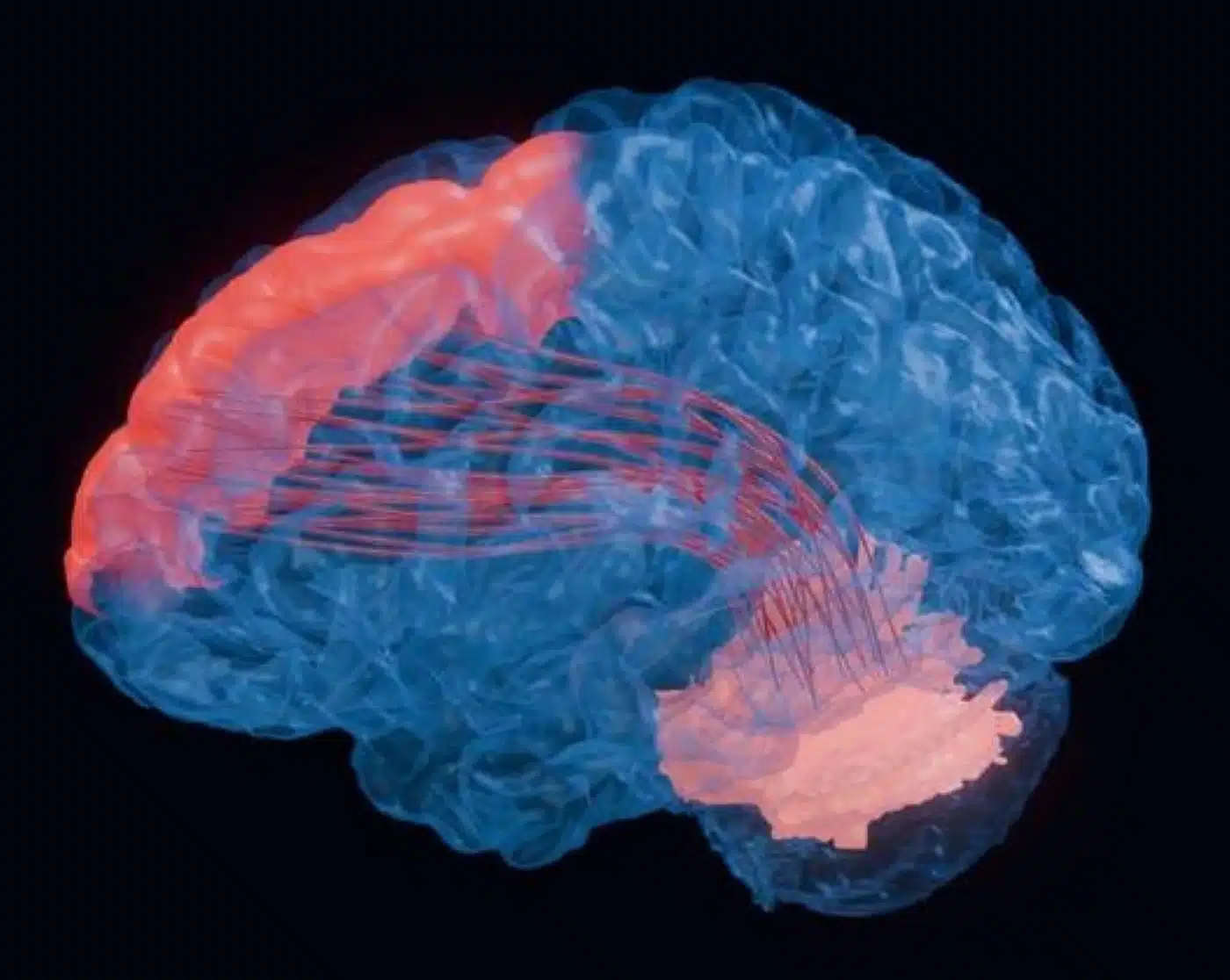
- Cerebrocerebellar Disconnection: Brain MRI analyses of PH cohorts revealed consistent tissue atrophy and weakened functional connectivity specifically linking the cerebellum to the prefrontal cortex—neural circuits essential for working memory, attention, and executive planning.
- Driven by the NCOA7 Gene Variant: The NCOA7 gene variant, which exacerbates vascular inflammation in approximately 25% of PH patients, was directly tied to cerebellar neuroinflammation; knockout mice lacking NCOA7 exhibited markedly exacerbated cerebellar microglial activation and neuronal remodeling.
- Independent of Systemic Hypoxia: Brain abnormalities were entirely specific to PH and could not be explained by low blood oxygen levels, cardiac strain, or pharmacological treatments, ruling out secondary hypoxic damage and establishing a distinct neuro-vascular inflammatory pathology.
Source: University of Pittsburgh / UPMC
Pulmonary hypertension (PH) is a progressive, life-threatening vascular disease characterized by pathologically elevated blood pressure within the pulmonary arterial bed, placing severe strain on the right ventricle of the heart. While classical clinical management focuses on cardiorespiratory symptoms, including exertional dyspnea, fatigue, angina, and syncope, a substantial proportion of patients report debilitating cognitive deficits, including memory lapses, impaired decision-making, and sluggish executive function.
For decades, clinicians frequently attributed these cognitive complaints to secondary complications: reduced cerebral oxygenation due to poor lung gas exchange, reduced cardiac output, or the side effects of complex vasodilator therapies.
Now, an investigation published in the Journal of Clinical Investigation by researcher-clinicians at the University of Pittsburgh School of Medicine and UPMC demonstrates that cognitive decline in pulmonary hypertension is a primary manifestation of the disease itself. The same inflammatory cascade that constricts and remodels the pulmonary vasculature also crosses organ systems to drive localized neuroinflammation and structural brain atrophy.
Mapping the Cerebrocerebellar Disconnection
The study was directed by Stephen Chan, MD, PhD, Vitalant Chair in Vascular Medicine and director of the Vascular Medicine Institute and Center for Pulmonary Vascular Biology and Medicine at Pitt and UPMC.
Chan’s team analyzed neuroimaging and biomarker profiles across two cohorts: a large population-level public health dataset and a specialized clinical cohort of PH patients treated at UPMC. Brain MRI scans uncovered a distinct anatomical signature:
- Specific Tissue Loss: Patients with pulmonary hypertension exhibited marked, consistent loss of brain parenchyma concentrated in the cerebellum and prefrontal regions.
- Circuit Breakdown: Functional connectivity analyses revealed disrupted communication along the cerebrocerebellar loop, the white-matter pathway through which the cerebellum coordinates and modulates frontal lobe cognitive processing, working memory, and executive planning.
Crucially, this structural and functional signature was absent in control cohorts diagnosed with other severe cardiovascular diseases, chronic pulmonary disorders, or psychiatric illnesses. Furthermore, multivariate statistical models established that the cerebellar tissue loss could not be accounted for by arterial oxygen desaturation, right ventricular failure, or disease-targeted medications, confirming that the damage represents a primary neuro-inflammatory process rather than systemic hypoxia.
“This changes the way we look at a disease like pulmonary hypertension,” said Dr. Chan. “In medicine, we typically treat the symptoms of one organ system; but the reality is that our bodies are very complex, and disease processes may be affecting multiple parts of the body in a variety of ways.”
The NCOA7 Inflammatory Cascade
In previous vascular biology research, Dr. Chan identified a functional genetic variant in NCOA7 (nuclear receptor coactivator 7). Present in approximately 25% of pulmonary hypertension patients, this variant fuels hyperinflammatory responses in blood vessels, accelerating arterial remodeling and worsening clinical mortality.
Hypothesizing that NCOA7 dysregulation extends into the central nervous system, the researchers evaluated postmortem cerebellar tissue from both human patients and preclinical murine models of PH:
- Microglial Activation: In the cerebellum of PH models, resident immune cells (microglia) adopted a hyperactive, pro-inflammatory morphology, triggering downstream synaptic and dendritic remodeling in neighboring neurons.
- Genetic Causality Confirmed: Preclinical mice engineered to develop pulmonary hypertension while lacking functional NCOA7 exhibited significantly more severe neuroinflammation and accelerated cerebellar tissue breakdown than wild-type PH controls, demonstrating that NCOA7 serves as a critical molecular brake against neuroinflammation.
Expanding the Paradigm Beyond the Lungs
Because NCOA7 regulates fundamental inflammatory pathways, the investigators believe this mechanism may influence a broader spectrum of systemic vascular and neurodegenerative disorders.
“I believe that NCOA7 deficiency may underlie a spectrum of inflammatory conditions, including those that may contribute to dementia and aging,” Dr. Chan stated. “If so, these findings could introduce new diagnostic and therapeutic avenues for preserving brain function in a much broader group of people.”
Currently, international clinical guidelines for pulmonary hypertension do not recommend routine cognitive screening, leaving many patients to struggle with unrecognized brain fog. Identifying NCOA7 and downstream microglial activation pathways provides a measurable biological marker to screen for future neurocognitive vulnerability, while offering a targeted pharmacological avenue to preserve cognitive function and overall quality of life.
“Patients often assume memory issues or trouble concentrating are unrelated to their pulmonary hypertension,” Dr. Chan added. “Our findings suggest those symptoms are a central part of the disease process and deserve attention.”
Funding: Funding for this work was provided by the National Institutes of Health (R35GM164224, R01HL 161038), American Heart Association (26SFRNPVHD1609644, 24SFRNPCN1280228, 24CDA1274310), National Aeronautics and Space Administration (80NSSC26K0720), WoodNext Foundation, Collaborative Research Center (1213 Project A10N, SA 1923/7-1, 1213 Project A01, A05), U.S. Department of Veterans Affairs (IK6 BX005647-01, I01BX006894-01), and the European Research Council Consolidator (866051).
Chan and Lloyd Harvey are inventors on patents and patent applications regarding metabolism and inflammation in pulmonary hypertension and pharmacologic targeting of NCOA7.
Editorial Notes:
- This article was edited by a Neuroscience News editor.
- Journal paper will be reviewed in full upon release.
- Additional context added by our staff.
About this neurology and cognition Research:
- Media Contact: Zachary Sweger
- Source: University of Pittsburgh
- Image Credit: Image credited to Satoshi Okawa, Ph.D.
- Original Research is Open Access: The findings will appear in Journal of Clinical Investigation


