Summary: A new study sheds light on how the primate brain evolved and intelligence developed.
Source: University of Florida.
New study sheds light on evolution of human, ape intelligence.
Virtual brains reconstructed from ancient, kiwi-sized primate skulls could help resolve one of the most intriguing evolutionary mysteries: how modern primates developed large brains.
University of Florida paleontologists found clues in the remarkably preserved skulls of adapiforms, lemur-like primates that scurried around the tropical forests of Wyoming about 50 million years ago. Thought to be a link between primitive and advanced primates, their fossil skulls were the best evidence available for understanding the neuroanatomy of the earliest ancestors of modern primates. But there was just one problem—the brain cavities of the fragile skulls contained only rock and dust.
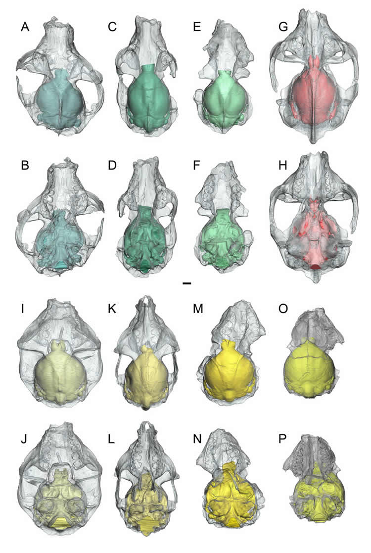
That is, until Arianna Harrington, then a UF undergraduate student and later a master’s student at the Florida Museum of Natural History on the UF campus, used CT technology to create the first virtual 3-D brain casts of the early primates. The eight virtually reconstructed and dissected brains—the most ever created for a single study—show an evolutionary burst including improved vision and more complex neurological function preceded an increase in brain size, said Harrington, now a Duke University doctoral student. Details of the findings are described online in the Journal of Human Evolution.
“It may be that these early specializations allowed primate brains to expand later in time,” said Harrington, the study’s lead author. “The idea is that any patterns we find in primate brain evolution could lead to a better understanding of the early evolution that led to the human brain.”
Scientists have long debated whether primates have always had big brains compared to body size, or if this was a trait that appeared later. The new study’s findings are consistent with previous endocast studies of Australopithecus afarensis, the oldest hominid known, and Victoriapithecus macinnesi, an early Old World monkey, which showed brain size increase followed brain specialization in early hominids and monkeys.
Adapiforms, which are not directly related to humans, evolved after the earliest primate ancestors, called plesiadapiforms, which lived about 65 million years ago. Harrington and colleagues created virtual endocasts for three different species of adapiforms: Notharctus tenebrosus and Smilodectes gracilis from the middle Eocene Bridger formation of Wyoming and a late Eocene European specimen named Adapis parisiensis.
Adapiforms’ skulls differ from the earlier plesiadapiforms in a few ways including having more forward-facing eyes. Thanks to the new virtual endocasts, scientists were able to take a closer look at anatomical features which revealed that, while adapiforms placed relatively less emphasis on smell more similar to modern primate brains, the relative brain size was not so different from that of plesiadapiforms, said study co-author Jonathan Bloch, curator of vertebrate paleontology at the Florida Museum.
“While it’s true humans and other modern primates have very large brains, that story started down at the base of our group,” Bloch said. “As our study shows, the earliest primates actually had relatively small brains. So they didn’t start out with large brains and maintain them.”
Modern primates are specialized in the visual sense. One of the main differences between the early plesiadapiforms and adapiforms is the region of the brain responsible for the sense of smell, the olfactory bulb, is smaller, while there appears to be an expansion in the area of the brains responsible for vision, Harrington said.
“It is likely this indicates they’re beginning to rely more on vision than smell,” she said. “Scientists have hypothesized that vision may have helped early primates forage in complex arboreal forest systems.”
Funding: The research was supported by the National Institutes of Health (GM0709077 and AI117911).
Source: University of Florida
Image Source: This NeuroscienceNews.com image is credited to University of Florida.
Original Research: Abstract for “First virtual endocasts of adapiform primates” by Arianna R. Harrington, Mary T. Silcox, Gabriel S. Yapuncich, Doug M. Boyer, and Jonathan I. Bloch in Journal of Human Evolution. Published online August 2016 doi:10.1016/j.jhevol.2016.06.005
[cbtabs][cbtab title=”MLA”]University of Florida. “How Did Primate Brains Get So Big?.” NeuroscienceNews. NeuroscienceNews, 11 August 2016.
<https://neurosciencenews.com/primate-brain-size-evolution-4833/>.[/cbtab][cbtab title=”APA”]University of Florida. (2016, August 11). How Did Primate Brains Get So Big?. NeuroscienceNews. Retrieved August 11, 2016 from https://neurosciencenews.com/primate-brain-size-evolution-4833/[/cbtab][cbtab title=”Chicago”]University of Florida. “How Did Primate Brains Get So Big?.” https://neurosciencenews.com/primate-brain-size-evolution-4833/ (accessed August 11, 2016).[/cbtab][/cbtabs]
Abstract
First virtual endocasts of adapiform primates
Well-preserved crania of notharctine adapiforms from the Eocene of North America provide the best direct evidence available for inferring neuroanatomy and encephalization in early euprimates (crown primates). Virtual endocasts of the notharctines Notharctus tenebrosus (n = 3) and Smilodectes gracilis (n = 4) from the middle Eocene Bridger formation of Wyoming, and the late Eocene European adapid adapiform Adapis parisiensis (n = 1), were reconstructed from high-resolution X-ray computed tomography (CT) data. While the three species share many neuroanatomical similarities differentiating them from plesiadapiforms (stem primates) and extant euprimates, our sample of N. tenebrosus displays more variation than that of S. gracilis, possibly related to differences in the patterns of cranial sexual dimorphism or within-lineage evolution. Body masses predicted from associated teeth suggest that N. tenebrosus was larger and had a lower encephalization quotient (EQ) than S. gracilis, despite their close relationship and similar inferred ecologies. Meanwhile, body masses predicted from cranial length of the same specimens suggest that the two species were more similar, with overlapping body mass and EQ, although S. gracilis exhibits a range of EQs shifted upwards relative to that of N. tenebrosus. While associated data from other parts of the skeleton are mostly lacking for specimens included in this study, measurements for unassociated postcrania attributed to these species yield body mass and EQ estimates that are also more similar to each other than those based on teeth. Regardless of the body mass prediction method used, results suggest that the average EQ of adapiforms was similar to that of plesiadapiforms, only overlapped the lower quadrant for the range of extant strepsirrhines, and did not overlap with the range of extant haplorhines. However, structural changes evident in these endocasts suggest that early euprimates relied more on vision than olfaction relative to plesiadapiforms, despite having relatively small endocranial volumes compared to extant taxa.
“First virtual endocasts of adapiform primates” by Arianna R. Harrington, Mary T. Silcox, Gabriel S. Yapuncich, Doug M. Boyer, and Jonathan I. Bloch in Journal of Human Evolution. Published online August 2016 doi:10.1016/j.jhevol.2016.06.005