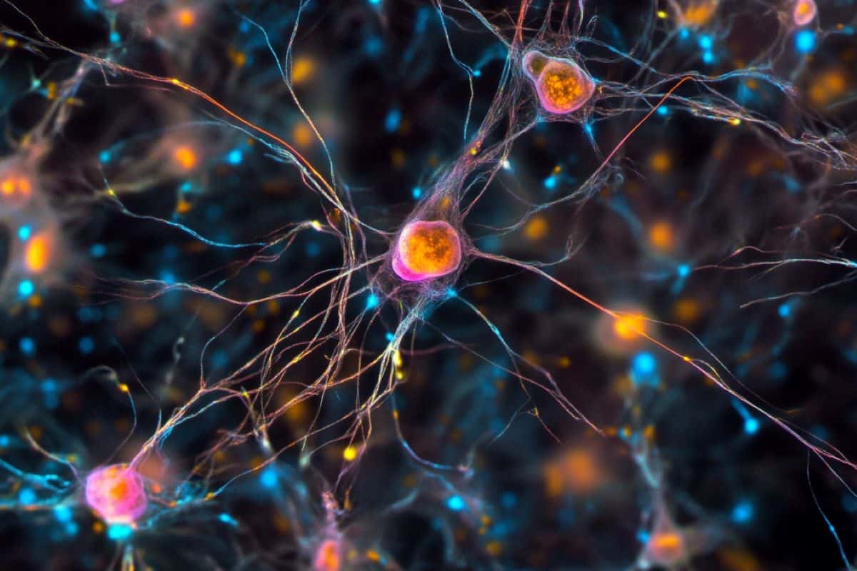
Summary: Researchers discovered that spontaneous brain activity during early development drives neural wiring before sensory experiences shape the brain. This spontaneous activity in neurons strengthens connections, following Hebb’s rule, where “cells that fire together wire together.”
The study focused on mouse retinal ganglion cells and found that synchronized activity leads to the growth of new neural branches, laying the groundwork for future learning and brain function. This finding sheds light on the fundamental processes guiding brain development and potential insights into neurological conditions.
Key facts
- Spontaneous neural activity in early development drives brain wiring.
- The study focused on mouse retinal cells before they experience sensory input.
- Synchronized activity leads to the growth of neural connections, following Hebb’s rule.
Source: Yale
In humans, the process of learning is driven by different groups of cells in the brain firing together. For instance, when the neurons associated with the process of recognizing a dog begin to fire in a coordinated manner in response to the cells that encode the features of a dog — four legs, fur, a tail, etc. — a young child will eventually be able to identify dogs going forward.
But brain wiring begins before humans are born, before they have experiences or senses like sight to guide this cellular circuitry. How does that happen?
In a new study published Aug. 15 in Science, Yale researchers identified how brain cells begin to coalesce into this wired network in early development before experience has a chance to shape the brain.

It turns out that very early development follows the same rules as later development — cells that fire together wire together. But rather than experience being the driving force, it’s spontaneous cellular activity.
“One of the fundamental questions we are pursuing is how the brain gets wired during development,” said Michael Crair, co-senior author of the study and the William Ziegler III Professor of Neuroscience at Yale School of Medicine. “What are the rules and mechanisms that govern brain wiring? These findings help answer that question.”
For the study, researchers focused on mouse retinal ganglion cells, which project from the retina to a region of the brain called the superior colliculus where they connect to downstream target neurons.
The researchers simultaneously measured the activity of a single retinal ganglion cell, the anatomical changes that occurred in that cell during development, and the activity of surrounding cells in awake neonatal mice whose eyes had not yet opened. This technically complex experiment was made possible by advanced microscopy techniques and fluorescent proteins that indicate cell activity and anatomical changes.
Previous research has shown that before sensory experience can take place — for instance, when humans are in the womb or, in the days before young mice open their eyes — spontaneously generated neuronal activity correlates and forms waves.
In the new study, researchers found that when the activity of a single retinal ganglion cell was highly synchronized with waves of spontaneous activity in surrounding cells, the single cell’s axon — the part of the cell that connects to other cells — grew new branches. When the activity was poorly synchronized, axon branches were instead eliminated.
“That tells us that when these cells fire together, associations are strengthened,” said Liang Liang, co-senior author of the study and an assistant professor of neuroscience at Yale School of Medicine. “The branching of axons allows more connections to be made between the retinal ganglion cell and the neurons sharing the synchronized activity in the superior colliculus circuit.”
This finding follows what’s known as “Hebb’s rule,” an idea put forward by psychologist Donald Hebb in 1949; at that time Hebb proposed that when one cell repeatedly causes another cell to fire, the connections between the two are strengthened.
“Hebb’s rule is applied quite a lot in psychology to explain the brain basis of learning,” said Crair, who is also the vice provost for research and a professor of ophthalmology and visual science. “Here we show that it also applies during early brain development with subcellular precision.”
In the new study, the researchers were also able to determine where on the cell branch formation was most likely to occur, a pattern that was disrupted when the researchers disturbed synchronization between the cell and the spontaneous waves.
Spontaneous activity occurs during development in several other neural circuits, including in the spinal cord, hippocampus, and cochlea. While the specific pattern of cellular activity would be different in each of those areas, similar rules may govern how cellular wiring takes place in those circuits, said Crair.
Going forward, the researchers will explore whether these patterns of axon branching persist after a mouse’s eyes open and what happens to the downstream connected neuron when a new axon branch forms.
“The Crair and Liang labs will continue to combine our expertise in brain development and single-cell imaging to examine how the assembly and refinement of brain circuits is guided by precise patterns of neural activity at different developmental stages,” said Liang.
Funding: The research was supported in part by the Kavli Institute of Neuroscience at Yale School of Medicine.
About this neurodevelopment research news
Author: Bess Connolly
Source: Yale
Contact: Bess Connolly – Yale
Image: The image is credited to Neuroscience News
Original Research: Closed access.
“Hebbian instruction of axonal connectivity by endogenous correlated spontaneous activity” by Michael Crair et al. Science
Abstract
Hebbian instruction of axonal connectivity by endogenous correlated spontaneous activity
INTRODUCTION
During the development of the mammalian central nervous system, circuit refinement depends critically on neuronal activity. Prior to the onset of sensory experience, the sensory periphery spontaneously generates spatiotemporal patterns of activity that synchronize the firing among neurons within and between brain areas.
These spontaneous neuronal activity patterns are required to establish the initial configuration of functional circuits necessary for early behavior and survival.
Although previous studies have demonstrated the critical role of synchronous activity on circuit connectivity, the enduring and intriguing question remains of how endogenously generated patterns of spontaneous activity instruct functional circuit refinement at the cellular and subcellular levels.
RATIONALE
To investigate the instructive role of spontaneous activity on fine-scale circuit refinement, we established a simultaneous in vivo two-photon imaging method that combines time-lapse imaging of a single axon with dual-color calcium imaging of both single-axon activity and activity in the surrounding population of axons or target neurons in the mouse superior colliculus.
During early postnatal development, spontaneous waves of patterned activity propagate in the developing retina and central nervous system to drive axon refinement before eye opening. With this multi-imaging method, we simultaneously recorded (i) single–retinal ganglion cell (RGC) axon branch dynamics [through enhanced green fluorescent protein (EGFP)], (ii) axon firing (through GCaMP6s, a genetically encoded calcium indicator) of the same EGFP-expressing single RGC axon, and (iii) patterns of population activity (through jRGECO1a, a red-shifted genetically encoded calcium indicator) among RGC axons or superior colliculus neurons (i.e., retinal waves) in awake, behaving mice.
RESULTS
With this experimental approach, we observed that individual axon branches in RGCs have different levels of synchronization with retinal waves at the subcellular level, resulting in a heterogeneous single-axon spatial correlation map between the firing activity of the axon and its neighbors.
The degree of activity synchronization predicts where an axon branch is added or eliminated: Axon branches are preferentially eliminated in regions with low levels of local synchronization between single-axon firing and retinal waves but added in regions with high levels of local synchronization.
The instructive role of retinal waves in axon branch dynamics was diminished by decoupling single-axon firing and retinal waves with sparse conditional knockout of nicotinic acetylcholine receptors (nAChRs) containing β2 subunits from individual RGC axons or by pharmacological blockade of N-methyl-d-aspartate receptors (NMDARs).
By combining in vivo single-axon glutamate imaging with time-lapse imaging of single-axon branch dynamics, we showed that axon branches were preferentially eliminated in regions where glutamate release sites (GRSs) are less dense and that new branches preferentially emerged from presynaptic sites with high levels of glutamate release.
CONCLUSION
Our findings demonstrate that endogenous patterns of spontaneous activity instruct axon branch addition and elimination with subcellular precision following Hebb’s predictions, colloquially described as “cells that fire together, wire together” and “out of sync, lose your link.”
Our observations further suggest that the instructive role of retinal waves in axon remodeling is mediated by synaptic plasticity: Patterned spontaneous activity stabilizes and strengthens individual presynaptic sites at the subcellular compartment level following Hebb’s law, and the positions and strength of presynaptic sites further determine where axon branches are added or eliminated.
Our results provide the first direct in vivo evidence that endogenous patterns of spontaneous activity drive circuit refinement through Hebbian plasticity rules and elucidate the activity-dependent mechanism for how functional brain circuits are self-organized even before the onset of sensory experience.


