Summary: Researchers report how a key protein in the blood coagulation pathway folds to a higher energy state.
Source: University of Massachusetts Amherst.
Protein-folding research at UMass Amherst reports for the first time how a key protein in the blood coagulation pathway folds to a higher-energy ‘cocked’ state
Though research on protein folding has progressed over the past few decades toward better understanding of human metabolism and the diseases associated with misfolding, important discoveries are still being made by teams who can bring special techniques and tools to bear on these complex cellular processes.
Today a research group with expertise in protein folding at the University of Massachusetts Amherst led by biochemist Daniel Hebert reports for the first time how a key protein in the blood coagulation pathway folds to a higher-energy or “cocked” state, so it can function as a sort of “molecular mousetrap” and generate the work required to perform physiologically important functions. Details appear in an early online edition of Proceedings of the National Academy of Sciences.
This work advances understanding of genetic forms of thrombosis, emphysema, cirrhosis of the liver, neurodegenerative diseases and inflammation, among others.
Hebert says, “Generally these proteins don’t have a lot of mobility, but this particular protein is folded like a spring-loaded trap, so it is stable at a higher energy state. When it’s needed, it fires from a loaded position and undergoes the big structural changes needed to carry out its inhibitory function against target proteases. We can explain now for the first time how it folds in live cells into an active state and how it avoids folding to a more stable but inactive state.”
He and biophysicist Lila Gierasch, with Anne Gershenson, a specialist in the serpin family of proteins and serinopathies they studied, used radio-labeled pulse-chase techniques to follow the serpin antithrombin III (ATIII) through time as it folded. They also used antibodies to isolate it and gel electrophoresis to analyze its expression, folding and function.
Hebert says it takes about one hour overall for cells to synthesize, fold and secrete the serpin ATIII, but he and colleagues found that in the critical first few minutes of this process a key positioning of the mousetrap’s trigger wire takes place, and the rest of the trap builds around it, allowing it to reach the loaded higher energy conformation.
“We found it a bit surprising that the part of the protein that is synthesized last is locked into place early in the folding process, helping to guide later structure formation. But on reflection it makes sense, because the amount of energy it would take to cock the trigger at the end would be prohibitive. So the protein starts out cocked, and goes on from there.”
The research team introduced a number of disease-associated mutations and watched where proteins containing these mutations diverged from normal folding. Their paper not only introduces and defines for the first time the early stages of the normal pathway but it describes how 13 mutations found in humans affect ATIII folding. They are already working with medical geneticists on identifying pharmacological chaperones to help mutated, misfolded proteins fold correctly and develop better constructs for gene therapies, Hebert says.
“We figured out the key for this protein to fold correctly,” he adds. “We utilized a type of serpin with a particular kind of covalent bond that would give us an experimental advantage that we could exploit to map the folding reaction. We chose this target deliberately and it allowed us to sort out the earliest stages that provided the key to how the spring gets loaded.”
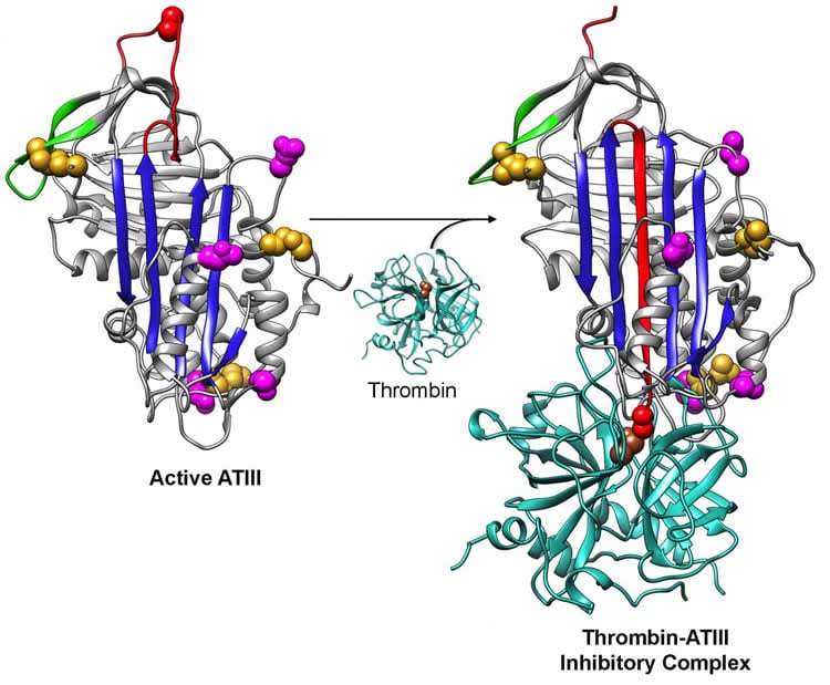
The serpin family is an important class of proteins that control diverse biological processes, and unlike other proteins they don’t always fold to their most stable state, Hebert notes. In fact, as noted, ATIII folds to a higher energy state, which is harder to get to and offers more chances for problems to arise during protein structure formation. Misfolded and partly folded serpins will never make it out of the cell, leading to lower protein levels in blood or to the build up of a toxic conformation, either of which is associated with disease.
The authors note that their data identify steps essential to ATIII folding and the findings “serve as a jumping off point to explore both in vitro and in vivo folding of related serpins.” Results highlight possible answers to questions about how serpins “successfully navigate their folding landscape to produce adequate amounts of functional metastable protein and avoid deleterious aggregation,” and suggest promising areas for future work.
Funding: This work was supported by a EUREKA grant from the National Institutes of Health Institute of General Medical Sciences.
Source: Janet Lathrop – University of Massachusetts Amherst
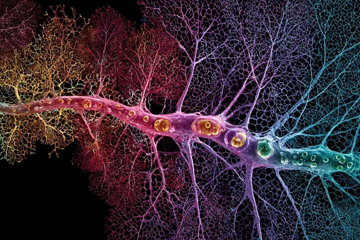
Image Source: This NeuroscienceNews.com image is adapted from the University of Massachusetts Amherst press release.
Original Research: The study will appear in PNAS.
[cbtabs][cbtab title=”MLA”]University of Massachusetts Amherst. “Untangling Disease Related Protein Misfolding.” NeuroscienceNews. NeuroscienceNews, 23 May 2016.
<https://neurosciencenews.com/neurology-protein-misfolding-4280/>.[/cbtab][cbtab title=”APA”]University of Massachusetts Amherst. (2016, May 23). Untangling Disease Related Protein Misfolding. NeuroscienceNews. Retrieved May 23, 2016 from https://neurosciencenews.com/neurology-protein-misfolding-4280/[/cbtab][cbtab title=”Chicago”]University of Massachusetts Amherst. “Untangling Disease Related Protein Misfolding.” https://neurosciencenews.com/neurology-protein-misfolding-4280/ (accessed May 23, 2016).[/cbtab][/cbtabs]
Abstract
Reminders Through Association
People often fail to follow through on good intentions. While limited self-control is frequently the culprit, another cause is simply forgetting to enact intentions when opportunities arise. We introduce a novel, potent approach to facilitating follow-through: the reminders-through-association approach. This approach involves associating intentions (e.g., to mail a letter on your desk tomorrow) with distinctive cues that will capture attention when you have opportunities to act on those intentions (e.g., Valentine’s Day flowers that arrived late yesterday, which are sitting on your desk). We showed that cue-based reminders are more potent when the cues they employ are distinctive relative to (a) other regularly encountered stimuli and (b) other stimuli encountered concurrently. Further, they can be more effective than written or electronic reminder messages, and they are undervalued and underused. The reminders-through-association approach, developed by integrating and expanding on past research on self-control, reminders, and prospective memory, can be a powerful tool for policymakers and individuals.
“Reminders Through Association” by Todd Rogers and Katherine L. Milkman in Psychological Science. Published online May 20 2016 doi:10.1177/0956797616643071