

Summary:
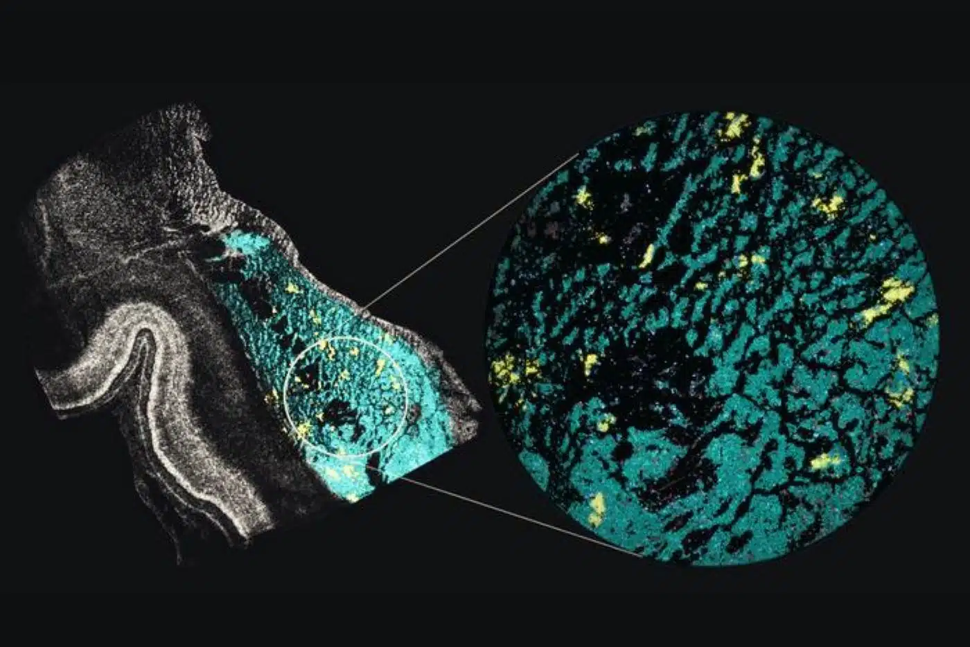
In a landmark study published in Cell, an NIH BRAIN Initiative consortium co-led by UCLA neuroscientists has mapped the molecular and 3D genomic development of the human basal ganglia from mid-pregnancy through infancy. Analyzing nearly 287,000 individual brain cells using advanced single-nucleus methyl-3C sequencing, researchers tracked how developing neurons migrate from temporary prenatal structures known as the ganglionic eminences.
The atlas revealed that spiny projection neurons mature in a highly coordinated manner early in gestation and carry strong enrichment for genetic variants linked to schizophrenia and bipolar disorder, establishing an unprecedented developmental reference to pinpoint when and where psychiatric risk takes root.
Key Facts:
- Mapping Epigenetic & 3D DNA Architecture: Using single-nucleus methyl-3C sequencing (snm3C-seq) across nearly 287,000 brain cells from mid-pregnancy through infancy, the team tracked both DNA methylation and 3D chromatin folding inside developing human cell nuclei.
- Divergent Maturation Timelines: Cells originating in temporary prenatal ganglionic eminences follow distinct developmental clocks, while some mature by mid-pregnancy, others continue generating immature neurons into infancy. Spiny projection neurons showed coordinated maturation and signs of electrical activity as early as mid-gestation.
- Pinpointing Psychiatric Risk Windows: Cross-referencing the developmental atlas with large-scale genome-wide association studies revealed that developing spiny projection neurons are strongly enriched for genetic variants associated with schizophrenia and bipolar disorder.
Source: UCLA / Cell
During fetal development, the human brain undergoes an extraordinary cellular migration. Newly born neurons arise in transient, specialized germinal zones and travel long distances across developing tissue to assemble into functional circuits.
Because many of these temporary prenatal structures vanish around the time of birth, capturing the molecular transformations that unfold during this migration has historically been one of neurobiology’s greatest technical challenges.
One critical destination for these migrating cells is the basal ganglia, a deep cluster of subcortical nuclei governing motor control, habit formation, reward processing, and behavioral regulation. Dysfunction within basal ganglia circuitry is implicated across a broad spectrum of neurological and psychiatric disorders, including Parkinson’s disease, Huntington’s disease, addiction, obsessive-compulsive disorder (OCD), and schizophrenia.
Now, in a study published in the journal Cell, a multi-institutional research team co-led by scientists at the University of California, Los Angeles (UCLA), UC San Francisco, and UC San Diego has mapped the developmental trajectory of the human basal ganglia from mid-pregnancy through infancy in unprecedented 3D molecular detail.
Conducted as part of the National Institutes of Health’s BRAIN Initiative Cell Atlas Network (BICAN), the study establishes a foundational reference atlas showing how gene regulation and physical DNA folding orchestrate human brain maturation—and how inherited genetic risk for psychiatric disorders maps onto specific developing cell types.
Tracing Cells From Temporary Prenatal Structures
While standard single-cell atlases primarily catalog which genes are actively transcribed, they often miss the upstream epigenetic mechanisms that dictate when and where genes are switched on or off.
To uncover these regulatory blueprints, co-senior authors Dr. Chongyuan Luo and Dr. Jason Ernst of UCLA, alongside co-leads Dr. Mercedes Paredes (UCSF) and Dr. Eran Mukamel (UCSD), focused on the ganglionic eminences—transient prenatal brain structures that generate the majority of the brain’s inhibitory interneurons as well as key projection neurons destined for the basal ganglia.
“Genetic studies can tell us that particular regions of the genome are associated with disease, but they often can’t tell us which cell type is affected or when during development that genetic risk becomes relevant,” explained Luo, associate professor of human genetics at the David Geffen School of Medicine at UCLA and member of the UCLA Broad Stem Cell Research Center. “This atlas gives us that cellular and developmental context.”
“Because these structures disappear around birth, adult brain studies miss an important early part of these neurons’ development,” added co-first author Matthew Heffel, a former doctoral researcher in Luo’s laboratory. “By sampling them directly, we can compare cells near where they originate with related cells after they have migrated to other parts of the brain.”
Deploying a specialized technique co-developed by Luo’s team called single-nucleus methyl-3C sequencing (snm3C-seq), the investigators simultaneously measured DNA methylation patterns and the physical three-dimensional folding of chromatin inside more than 155,000 individual cell nuclei collected from nine developing human brains spanning mid-pregnancy to seven months postnatally. Integrating these profiles with existing developmental and adult datasets brought the total analytical atlas to nearly 287,000 cells.
Coordinated Spiny Projection Neurons and Myelin Reorganization
The expanded single-cell atlas revealed that neural populations arising from distinct subregions of the ganglionic eminences mature on sharply divergent biological schedules. While certain neuronal lineages achieved relative molecular maturity by mid-pregnancy, other progenitor pools continued generating immature neurons well into postnatal infancy.
Among these populations, spiny projection neurons, the primary output neurons of the basal ganglia, stood out. Unlike neighboring neural subtypes, spiny projection neurons matured in a highly synchronized, coordinated wave, exhibiting molecular signatures of functional neuronal activity as early as mid-pregnancy. This early activation suggests they actively participate in wiring basal ganglia circuits long before birth.
Postnatally, the atlas also captured a dramatic genomic restructuring event within oligodendrocytes—the glial support cells responsible for wrapping nerve fibers in insulating myelin sheaths. As immature oligodendrocyte progenitor cells differentiated into mature myelinating oligodendrocytes after birth, their nuclear DNA temporarily folded into a distinct 3D chromatin configuration directly linked to heightened transcriptional activity.
Connecting Prenatal Development to Schizophrenia Risk
To translate this basic developmental map into clinical insight, the researchers integrated their 3D epigenomic atlas with large-scale genome-wide association studies (GWAS) of major psychiatric disorders.
The analysis demonstrated that developing spiny projection neurons harbor an exceptionally strong enrichment for genetic risk variants tied to schizophrenia, with specific subtypes also showing significant associations with bipolar disorder.
Because the study evaluated neurotypical developing tissue rather than patient samples, the findings do not indicate that these neurons directly cause schizophrenia. Rather, the atlas narrows down the exact cellular and developmental windows during which inherited genetic variations may alter circuit assembly.
“Researchers can compare what they see in patients or disease models with typical development to identify the cell types and molecular features that may be most relevant to disease,” said co-senior author Jason Ernst, UCLA professor of biological chemistry, computer science, and computational medicine.
By providing an open, high-resolution epigenetic and 3D genomic reference of human brain assembly, the BICAN basal ganglia atlas equips neuroscientists with a vital tool to decode the developmental origins of neuropsychiatric illness.
Questions and Answers
What is the basal ganglia, and why did researchers map its development?
The basal ganglia is a group of deep brain structures essential for controlling movement, habits, reward, and behavior, and it is heavily implicated in conditions like Parkinson’s disease, addiction, OCD, and schizophrenia. Researchers mapped its development from mid-pregnancy to infancy to capture critical molecular changes in transient prenatal brain structures that disappear around birth.
How did this study differ from previous brain cell atlases?
While most brain atlases simply measure which genes are turned on or off, this study used single-nucleus methyl-3C sequencing (snm3C-seq) across nearly 287,000 cells to simultaneously map DNA methylation and how DNA physically folds in 3D space inside individual cell nuclei, revealing how gene regulation is controlled during cell migration and maturation.
How does this atlas connect to schizophrenia and bipolar disorder?
When researchers overlaid genetic risk variants from large psychiatric studies onto their developmental map, they discovered that developing spiny projection neurons in the basal ganglia were strongly enriched for genetic variants linked to schizophrenia and bipolar disorder, helping pinpoint when and where inherited risk acts during early brain assembly.
Funding: The study was co-led by Dr. Mercedes Paredes of UC San Francisco and Eran Mukamel of UC San Diego and was funded by the NIH, with additional support from the Chan Zuckerberg Initiative.
Editorial Notes:
- This article was edited by a Neuroscience News editor.
- Journal paper reviewed in full.
- Additional context added by our staff.
About this brain mapping and schizophrenia Research:
- Media Contact: Ani Vahradyan
- Source: UCLA
- Image Credit: Image credited to Mohammad S. Baig, Luo Lab
- Original Research is Open Access: Cell (Nov 12, 2026). “A single-cell and spatial 3D multi-omic atlas of developing human basal ganglia.” Authors: Matthew G. Heffel, Heng Xu, Oier Pastor-Alonso, Xinzhe Li, Mohammad S. Baig, Rayyan Irfan Ghoor, Runjia Li, Colin Kern, Joonho Kum, Yi Zhang, Jon Paino, Gaurav Singh Rathore, Min Jen Tsai, Chu-Yi Tai, Grant Tucker, Zoey Zhao, Angie Hou, Zachary von Behren, Mohini Bhade, Siqian Li, Kadellyn Sandoval, Jessica Scholes, Felicia Codrea, Jeffrey Calimlim, Emma K. Liao, Gwyneth Leung, JaeYeon Kim, Alexandra LeFevre, Robert Johnson, Thomas Blanchard, Eleazar Eskin, Jonathan Flint, Jennifer A. Cotter, Bogdan Pasaniuc, Bogdan Bintu, Quan Zhu, Eran A. Mukamel, Jason Ernst, Mercedes F. Paredes, and Chongyuan Luo.
- DOI: 10.1016/j.cell.2026.10.008
Abstract
A single-cell and spatial 3D multi-omic atlas of developing human basal ganglia
Human basal ganglia (BG) neurons and inhibitory interneurons originate in the ganglionic eminences (GEs), transient structures in the ventral prenatal brain.
This study delineates the specification and maturation of GEs and GE-derived neurons using single-nucleus methyl-3C sequencing (snm3C-seq), spatial transcriptomics, and single-molecule chromatin + RNA imaging, and we found heterogeneous temporal progression across GE subregions.
Spiny projection neurons (SPNs), the principal cells of the BG, acquire distinct subtypes through synchronized 3D-epigenomic remodeling, in contrast to the asynchronous maturation of other neuron types.
Oligodendrocyte differentiation involves a transient short-range chromatin state during infancy, showing that the short-range and long-range chromatin conformations previously linked to terminal glial and neuronal fates can also be transient. The temporal cortex matured epigenomically faster than other cortical regions during this period.
Lastly, SPNs were enriched in genetic risk for neuropsychiatric disease. Our study extends findings on unique 3D-epigenomic dynamics during human brain development.


