Summary: New research reveals that children with autism have varying neuron densities in certain brain regions compared to their peers. Using advanced imaging from the ABCD study, scientists observed lower neuron density in regions associated with memory and problem-solving, while regions like the amygdala showed higher neuron density.
These differences were specific to autism and did not appear in children with other psychiatric conditions, indicating a distinct neurological profile. These findings could enhance our understanding of autism’s development and potentially guide targeted therapies for children on the spectrum.
Key Facts
- Children with autism show neuron density differences in specific brain regions.
- Lower neuron density was observed in areas tied to memory and reasoning.
- Findings were unique to autism, not present in other psychiatric conditions.
Source: University of Rochester
There is new evidence that the cells responsible for communication in the brain may be structured differently in children with autism.
Researchers at the Del Monte Institute for Neuroscience at the University of Rochester discovered that in some areas of the brain neuron density varies in children with autism when compared to the general population.
“We’ve spent many years describing the larger characteristics of brain regions, such as thickness, volume, and curvature,” said Zachary Christensen, MD/PhD candidate at the University of Rochester School of Medicine and Dentistry, and first author of the paper out today in Autism Research.

“However, newer techniques in the field of neuroimaging for characterizing cells using MRI, unveil new levels of complexity throughout development.”
Imaging provides new insight into brain development
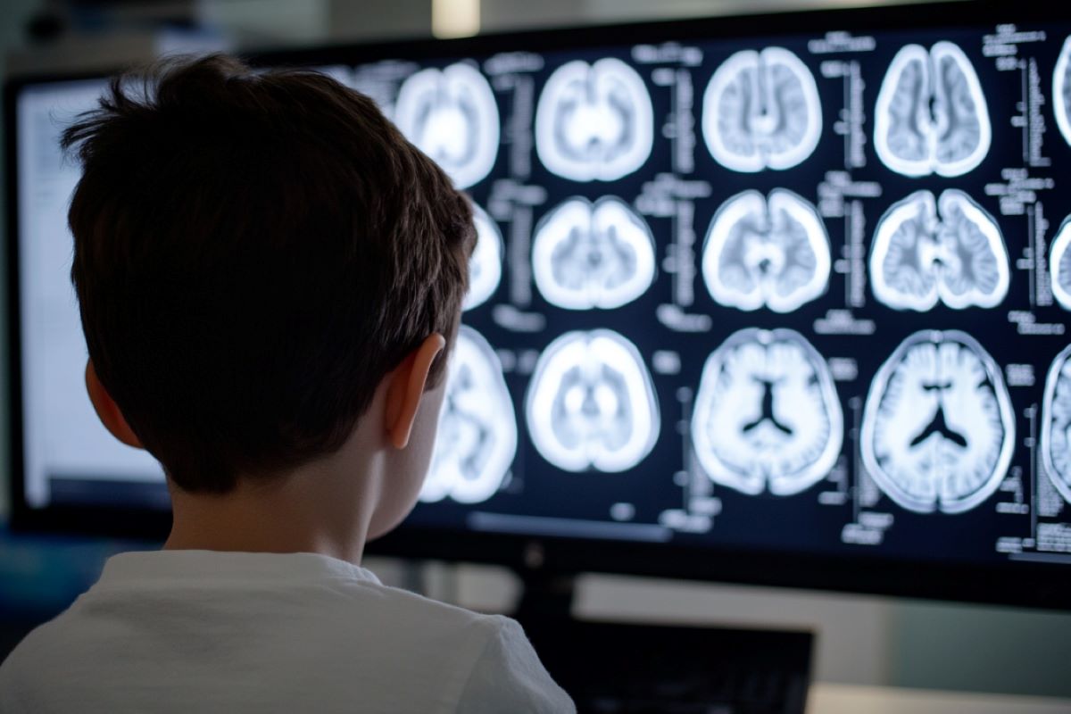
Researchers used brain imaging data collected from more than 11,000 children ages 9-11. They compared the imaging of the 142 children in that group with autism, to the general population and found there was lower neuron density in regions of the cerebral cortex. Some of these regions of the brain are responsible for tasks like memory, learning, reasoning, and problem-solving.
In contrast, the researchers also found other brain regions, such as the amygdala—an area responsible for emotions—that showed increased neuron density.
In addition to comparing the scans of children with autism to those of children without any neurodevelopmental diagnosis, they also compared the children with autism to a large group of children diagnosed with common psychiatric disorders like ADHD and anxiety.
The results were the same, suggesting that these differences are specific to Autism.
“People with a diagnosis of autism often have other things they have to deal with, such as anxiety, depression, and ADHD. But these findings mean we now have a new set of measurements that have shown unique promise in characterizing individuals with autism,” Christensen said.
“If characterizing unique deviations in neuron structure in those with autism can be done reliably and with relative ease, that opens a lot of opportunities to characterize how autism develops, and these measures may be used to identify individuals with autism that could benefit from more specific therapeutic interventions.”
Technology leverages what we know about the innerworkings of the brain and autism
Technology has transformed the level of precision and detail that investigators are now able to able to see in neuronal structure. Previously, researchers would only be able to see structural differences in neural populations postmortem.
The imaging data used for this research were collected from the Adolescent Brain Cognitive Development (ABCD) study database. It is the largest long-term study of brain development and child health.
The University of Rochester is one of 21 national sites collecting data for this study that began in 2015 and has revolutionized our understanding of adolescent brain health and development.
“We are at the beginning of understanding the true impact that the extraordinary data collected by the ABCD Study will have on the health of our children,” said John Foxe, PhD, senior author of the study, director of the Del Monte Institute for Neuroscience and the Golisano Intellectual and Developmental Disabilities Institute.
“It is truly transforming what we know about brain development as we follow this group of children from childhood into early adulthood.”
Additional authors include Edward Freedman of the University of Rochester Medical Center.
Funding: This research was supported by the Adolescent Brain Cognitive Development (ABCD) Study and the Translational Neuroimaging and Neurophysiology Core of the University of Rochester Intellectual and Developmental Disabilities Research Center.
About this Autism and neuroscience research news
Author: Kelsie Smith Hayduk
Source: University of Rochester
Contact: Kelsie Smith Hayduk – University of Rochester
Image: The image is credited to Neuroscience News
Original Research: Open access.
“Autism is associated with in vivo changes in gray matter neurite architecture” by Zachary Christensen et al. Autism Research
Abstract
Autism is associated with in vivo changes in gray matter neurite architecture
Postmortem investigations in autism have identified anomalies in neural cytoarchitecture across limbic, cerebellar, and neocortical networks. These anomalies include narrow cell mini-columns and variable neuron density.
However, difficulty obtaining sufficient post-mortem samples has often prevented investigations from converging on reproducible measures.
Recent advances in processing magnetic resonance diffusion weighted images (DWI) make in vivo characterization of neuronal cytoarchitecture a potential alternative to post-mortem studies.
Using extensive DWI data from the Adolescent Brain Cognitive Developmentsm (ABCD®) study 142 individuals with an autism diagnosis were compared with 8971 controls using a restriction spectrum imaging (RSI) framework that characterized total neurite density (TND), its component restricted normalized directional diffusion (RND), and restricted normalized isotropic diffusion (RNI).
A significant decrease in TND was observed in autism in the right cerebellar cortex (β = −0.005, SE =0.0015, p = 0.0267), with significant decreases in RNI and significant increases in RND found diffusely throughout posterior and anterior aspects of the brain, respectively.
Furthermore, these regions remained significant in post-hoc analysis when the autism sample was compared against a subset of 1404 individuals with other psychiatric conditions (pulled from the original 8971).
These findings highlight the importance of characterizing neuron cytoarchitecture in autism and the significance of their incorporation as physiological covariates in future studies.


