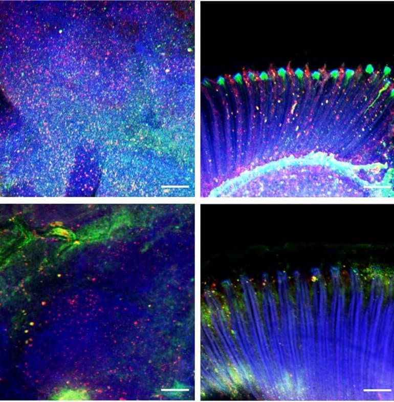
Summary: Mimicking a muscular stress system can provide neuroprotection against aging in both the brain and retina. The signal helps prevent the buildup of misfolded protein aggregates.
Source: St. Jude Children’s Research Hospital
How do different parts of the body communicate? Scientists at St. Jude are studying how signals sent from skeletal muscle affect the brain.
The team studied fruit flies and cutting-edge brain cell models called organoids. They focused on the signals muscles send when stressed. The researchers found that stress signals rely on an enzyme called Amyrel amylase and its product, the disaccharide maltose.
The scientists showed that mimicking the stress signals can protect the brain and retina from aging. The signals work by preventing the buildup of misfolded protein aggregates. Findings suggest that tailoring this signaling may potentially help combat neurodegenerative conditions like age-related dementia and Alzheimer’s disease.

“We found that a stress response induced in muscle could impact not only the muscle but also promote protein quality control in distant tissues like the brain and retina,” said Fabio Demontis, PhD, of St. Jude Developmental Neurobiology. “This stress response was actually protecting those tissues during aging.”
About this dementia research news
Source: St. Jude Children’s Research Hospital
Contact: Katy Hobgood – St. Jude Children’s Research Hospital
Image: The image is credited to St. Jude Children’s Research Hospital
Original Research: Closed access.
“Proteasome stress in skeletal muscle mounts a long-range protective response that delays retinal and brain aging” by Fabio Demontis et al. Cell Metabolism
Abstract
Proteasome stress in skeletal muscle mounts a long-range protective response that delays retinal and brain aging
Neurodegeneration in the central nervous system (CNS) is a defining feature of organismal aging that is influenced by peripheral tissues. Clinical observations indicate that skeletal muscle influences CNS aging, but the underlying muscle-to-brain signaling remains unexplored.
In Drosophila, we find that moderate perturbation of the proteasome in skeletal muscle induces compensatory preservation of CNS proteostasis during aging. Such long-range stress signaling depends on muscle-secreted Amyrel amylase.
Mimicking stress-induced Amyrel upregulation in muscle reduces age-related accumulation of poly-ubiquitinated proteins in the brain and retina via chaperones. Preservation of proteostasis stems from the disaccharide maltose, which is produced via Amyrel amylase activity. Correspondingly, RNAi for SLC45 maltose transporters reduces expression of Amyrel-induced chaperones and worsens brain proteostasis during aging.
Moreover, maltose preserves proteostasis and neuronal activity in human brain organoids challenged by thermal stress. Thus, proteasome stress in skeletal muscle hinders retinal and brain aging by mounting an adaptive response via amylase/maltose.


