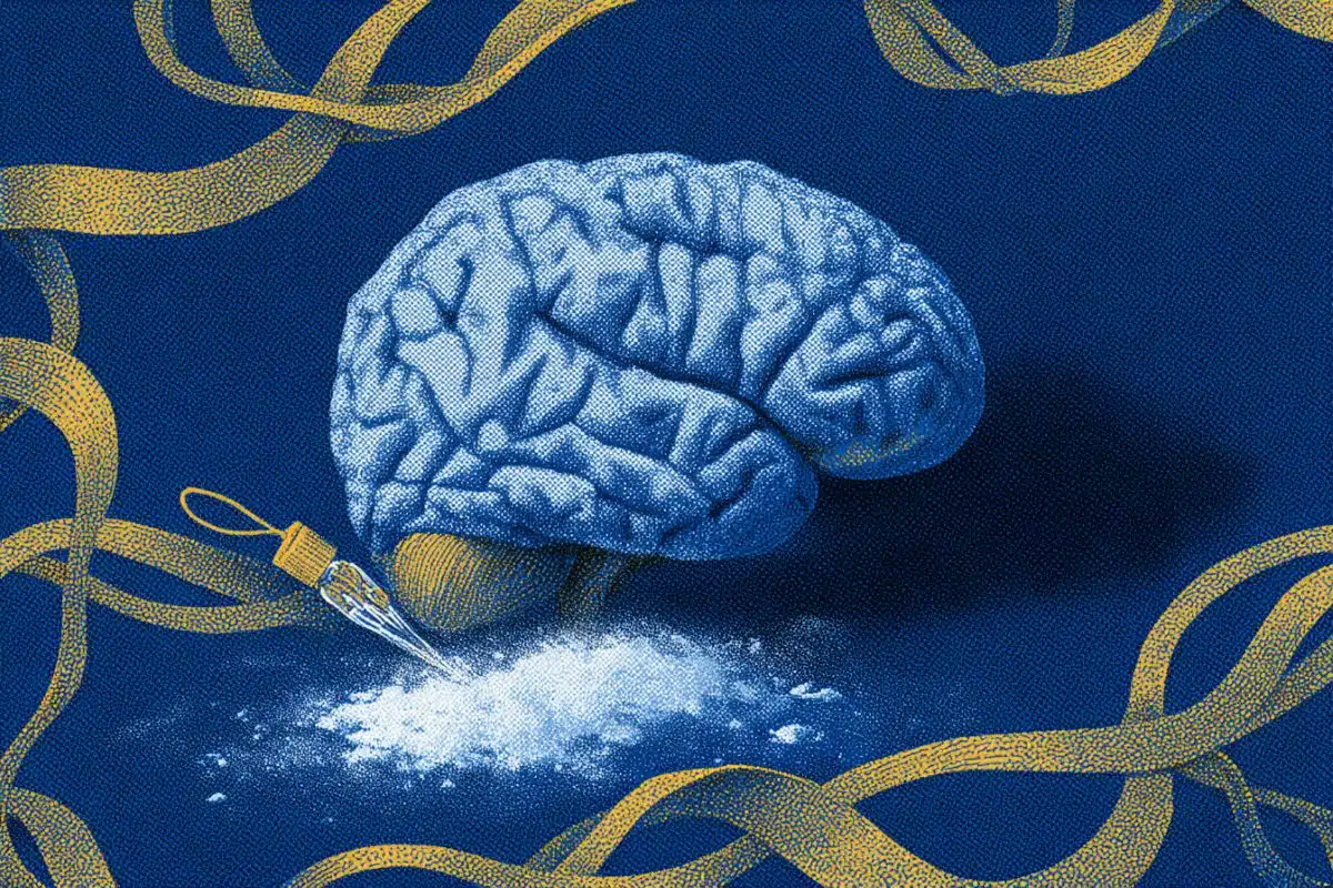
Summary: New research has revealed that myelin swellings—the precursors to lesions in Multiple Sclerosis (MS)—are not permanent fixtures of brain damage but are highly dynamic structures. Using advanced 3D microscopy, scientists discovered that these swellings can grow, shrink, and even disappear entirely.
Crucially, the study found that the activity level of the underlying nerve fiber dictates this process: high activity triggers larger swellings, while reduced activity can lead to total recovery. This shift in understanding suggests that the earliest stages of MS damage may be reversible, opening a massive new window for preventative treatments before protective myelin is permanently lost.
Key Facts
- Reversible Damage: Unlike previously thought, myelin swellings are dynamic; they have the capacity to shrink and recover completely rather than only progressing toward lesions.
- The Activity Link: The size and frequency of swellings are directly tied to the electrical activity of the nerve fiber; more active fibers experience more significant swelling.
- Advanced 3D Imaging: By using Third-Harmonic Generation (THG) and two-photon microscopy, researchers were able to observe these changes in three dimensions over time, moving past the limitations of traditional “frozen” tissue samples.
Source: KNAW
An international research team of Amsterdam UMC, VU LaserLab, the Netherlands Institute for Neuroscience and the University of Edinburgh have gained new insights into the dynamics of myelin swellings in the brain.
Myelin swellings are considered as the precursor of lesions in the brain of people with multiple sclerosis (MS).
The results have been recently published in the leading magazine Science.

MS is characterised by lesions in the brain and the spinal cord. Aside from these inflammations, damage can also be visible in the myelin; the protective layer surrounding nerve fibers. Myelin swellings are seen as a precursor for damaged myelin.
Dynamic damage
The research team used advanced microscopy techniques and different models – from zebrafish and mouse models to human brain tissue – to research the formation of this damage. This led to the discovery that myelin swellings have a dynamic character: they can not only grow, but also shrink and even recover completely.
It also turns out that the activity of the underlying nerve fibre plays an important role; more activity of the nerve fibre leads to more and bigger swellings, while less activity allows for possible recovery.
Advanced models
The strength of this study lies in the combination of different models and techniques, giving the researchers the opportunity to study the processes in both animal models and human tissue in an identical way. In traditional brain research, the tissue structure is usually frozen with chemicals, making it impossible to determine the dynamic nature of the myelin swelling.
In the collaboration between the laser lab and Amsterdam UMC, third-harmonic generation (THG) microscopy was used, while two-photon microscopy was performed at the Netherlands Institute for Neuroscience. Using these microscopy techniques, myelin changes in tissue could be studied in three dimensions and over time.
Start of myelin swellings
The next step is to explore why the activity of the nerve fibres can change the size and amount of myelin swellings. The researchers also want to study the role of brain cells in the appearance and disappearance of myelin swellings.
Using the published model systems, the team of Maarten Kole (NIN) and Antonio Luchicchi (MS Center Amsterdam, Amsterdam UMC), together with the team of David Lyons (Edinburgh), will seek to address these questions.
Addressing this early damage, even before myelin is lost, could offer in the future new treatment possibilities for MS and help keep myelin healthy for longer.
Source: Science
Funding: This research is made possible by MS Centrum Amsterdam, Nationaal MS Fonds, Progressive MS Alliance, The Friends Foundation of the Netherlands Institute for Neuroscience and NWO
Key Questions Answered:
A: Yes. The study highlights that the very first signs of damage—myelin swellings—are not a “point of no return.” Because they can shrink or vanish depending on nerve activity, there is potential to develop therapies that intervene during this window of recovery.
A: While the exact “why” is the next phase of research, the study clearly showed that the more a nerve fiber fires, the more the surrounding myelin swells. Finding a way to modulate this activity or the brain’s response to it could be the key to keeping myelin healthy.
A: Standard brain research often involves “fixing” tissue with chemicals, which effectively takes a static snapshot. By using advanced laser microscopy, this team was able to watch the myelin move and react in real-time, revealing a “lifecycle” of swelling that was previously invisible.
Editorial Notes:
- This article was edited by a Neuroscience News editor.
- Journal paper reviewed in full.
- Additional context added by our staff.
About this neurology and myelin research news
Author: Eline Feenstra
Source: KNAW
Contact: Eline Feenstra – KNAW
Image: The image is credited to Science
Original Research: Closed access.
“Myelin sheaths in the central nervous system can withstand damage and dynamically remodel” by Donia Arafa, Julia van de Korput, Philipp N. Braaker, Kieran P. Higgins, Niels R. C. Meijns, Katy L. H. Marshall-Phelps, Julia Meng, Daniel Soong, Eleonora Scalia, Kyle Lathem, Marcus Keatinge, Claire Richmond, Anna Klingseisen, Marja Main, Sarah A. Neely, David W. Hampton, Greg J. Duncan, Geert J. Schenk, Marie Louise Groot, Siddharthan Chandran, Ben Emery, Antonio Luchicchi, Maarten H. P. Kole, Anna C. Williams, and David A. Lyons. Science
DOI:10.1126/science.adr4661
Abstract
Myelin sheaths in the central nervous system can withstand damage and dynamically remodel
INTRODUCTION
Myelin is essential for normal central nervous system (CNS) function and is disrupted and/or lost in a range of human diseases. In conditions such as multiple sclerosis (MS) and in animal models of demyelination, the loss of myelin can trigger the regenerative process of remyelination, which is primarily mediated by the generation of new oligodendrocytes.
Although much is known about oligodendrocyte generation, myelin formation, and regeneration, we know far less about how myelin responds to damage. For example, it has remained unclear whether damaged myelin sheaths are lost in a similar manner irrespective of insult and to what extent damage necessarily leads to sheath loss.
It has also remained unknown whether damaged myelin sheaths of the CNS might even have the capacity to undergo repair.
RATIONALE
Motivated by evidence of its adaptability and potential for structural remodeling in the healthy nervous system, we investigated whether myelin in the CNS can withstand damage and possibly recover instead of being lost.
We reasoned that if myelin does indeed have a capacity to endure damage and dynamically remodel, then this might create new opportunities to protect myelin from loss and/or to promote its repair in human diseases.
RESULTS
Using a variety of zebrafish and rodent demyelination models in which damage was induced in distinct ways, we identified myelin swelling as an early hallmark of myelin damage that preceded overt myelin loss.
Using longitudinal live imaging in zebrafish and a rodent organotypic cortical slice culture model, we found that myelin sheath swelling did not necessarily prefigure myelin loss and that damage to sheaths could resolve over time. These observations indicated that damaged myelin has a capacity for dynamic structural remodeling.
Given that cell and tissue swelling is driven by disruption to ion and fluid homeostasis, we investigated whether neuronal activity might influence demyelination. Through a range of behavioral stimulation, optogenetic activation, and pharmacological interventions, we found that increasing neuronal activity exacerbated myelin swelling and decreased oligodendrocyte survival in zebrafish.
Conversely, reducing neuronal activity significantly mitigated myelin swelling in both zebrafish and mammalian slice models. Together, these observations indicate that neuronal activity represents a risk factor that can exacerbate early myelin pathology.
Extending these findings to humans, we analyzed postmortem MS tissue and found that swellings were prevalent in active and chronic active lesions, suggesting that myelin swelling represents an evolutionarily conserved feature of demyelination.
Live-imaging myelin pathology in acute postmortem MS tissue using high-resolution third harmonic generation imaging further revealed that myelin swelling could change dynamically over time and show signs of resolution in a human context.
CONCLUSION
Our findings demonstrate that early myelin damage, which is characterized by myelin swelling, is highly dynamic across species, including through human disease (MS). Our study reveals that damaged myelin has a capacity to remodel, which may represent an evolutionarily conserved mechanism to protect acutely compromised myelin from loss.
Targeting such early damage before myelin is lost may offer new therapeutic avenues for demyelinating disorders and for preserving myelin integrity with age.